
Description
Specifications
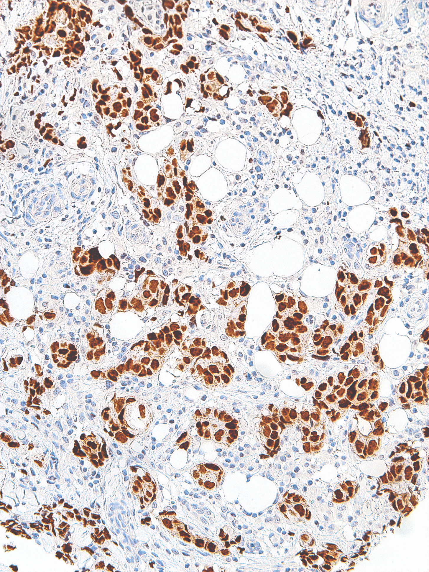
| Clone | IHC583 |
| Source | Mouse Monoclonal |
| Positive Control | Breast Carcinoma, Urothelial Carcinoma |
| Dilution Range | 1:200 |

| Clone | IHC583 |
| Source | Mouse Monoclonal |
| Positive Control | Breast Carcinoma, Urothelial Carcinoma |
| Dilution Range | 1:200 |

The MagPure Plasmid purification system uses the paramagnetic bead technology for high-throughput preparation of high-copy or low-copy plasmid DNA from E. coli cells. This kit also can be used with fosmid and BAC vector-based constructs. The system uses alkaline lysis followed by a MagPure purification to differentially bind plasmid DNA to paramagnetic beads. While the DNA is bound to the beads, contaminants can be rinsed away using a simple washing procedure. Because MagPure uses magnetic separation technology, the protocol does not require vacuum filtration. This makes kit extremely amenable to automation. Plasmid DNA purified with this system is most commonly used in Sanger Sequencing and PCR amplification.
Specifications
| Features | Specifications |
| Main Functions | Isolation up to 20µg plasmid DNA from 1-3ml bacterial culture |
| Applications | Enzyme digestion, sequencing, PCR and labeling, etc. |
| Purification technology | Magnetic beads technology |
| Process method | Manual or automatic |
| Sample type | Conventional plasmid, plasmid≤30KB |
| Sample amount | 1-3ml |
| Elution volume | ≥50μl |
This product is based on the purification method of high binding magnetic particles. The sample is lysed and digested under the action of lysate and Lysozyme. DNA is released into the lysate. After adding magnetic particles and binding solution, DNA will be adsorbed on the surface of magnetic particles, and impurities such as proteins will be removed without adsorption.The adsorbed particles were washed with washing solution to remove proteins and impurities, washed with ethanol to remove salts, and finally DNA was eluted by Elution Buffer.
Advantages
1. Suitable for extracting plasmids from 1-5ml or <3ml YT medium.
2. The same amount of buffer 1, 2, and 3 avoids errors caused by adjusting the pipette, making it convenient to use in conjunction with automated workstations.
3. Containing buffer 1 for washing, reducing the problem of false high production.
4. The purified plasmid can be directly used for sequencing, enzyme digestion, PCR, and other applications.
Kit Contents
| Contents | P181102 | P181103 | P181104 |
| Purification Times | 100 Preps | 500 Preps | 5000 Preps |
| RNase A | 10 mg | 50 mg | 2 x 250 mg |
| Buffer P1 | 30 ml | 150 ml | 2 x 750 ml |
| Buffer P2 | 30 ml | 150 ml | 2 x 750 ml |
| Buffer N3 | 30 ml | 150 ml | 2 x 750 ml |
| Buffer PW1 | 35 ml | 180 ml | 2 x 900 ml |
| MagPure Particle NB* | 2.2 ml | 11 ml | 2 x 60 ml |
Storage and Stability
RNase A and MagPure Particle NB should be stored at 2-8°C upon arrival. However, short-term storage (up to 24 weeks) at room temperature (15-25°C) does not affect its performance. The remaining kit components can be stored dry at room temperature (15-25°C) and are stable for at least 18 months under these conditions. After addition of RNase A, Buffer P1 is stable for 6 months when stored at 2-8°C.
Experiment Data

The MagPure Plasmid purification system uses the paramagnetic bead technology for high-throughput preparation of high-copy or low-copy plasmid DNA from E. coli cells. This kit also can be used with fosmid and BAC vector-based constructs. The system uses alkaline lysis followed by a MagPure purification to differentially bind plasmid DNA to paramagnetic beads. While the DNA is bound to the beads, contaminants can be rinsed away using a simple washing procedure. Because MagPure uses magnetic separation technology, the protocol does not require vacuum filtration. This makes kit extremely amenable to automation. Plasmid DNA purified with this system is most commonly used in Sanger Sequencing and PCR amplification.

Methyltetrazine-DBCO is a TCO reactive reagent containing a methyltetrazine group and a DBCO moiety. DBCO is commonly used for copper-free Click Chemistry reactions. Reagent grade, for research purpose. Please contact us for GMP-grade inquiries.

Methyltetrazine-DBCO is a TCO reactive reagent containing a methyltetrazine group and a DBCO moiety. DBCO is commonly used for copper-free Click Chemistry reactions. Reagent grade, for research purpose. Please contact us for GMP-grade inquiries.

Norgen’s Low Abundance DNA Quantification Kit offers a PCR-based detection procedure to quantify DNA of a wide spectrum of concentrations, including the lower ng per µL, pg per µL and sub-pg per µL range. The kit consists of a specially designed primer mix, that is used in conjunction with the provided 2x PCR Master Mix, to amplify human DNA from different types of inputs (such as various liquid biopsies). The kit is compatible with any Real-Time PCR system with the addition of fluorescent nucleic acid stains such as SYBR Green. The unknown DNA is accurately quantified by using a standard curve constructed from the provided DNA Standard.
Figure 1 / 2
Click for expanded view
Storage Conditions
Upon receipt, store Norgen’s Low Abundance DNA Quantification Kit at -20°C or lower. Avoid multiple freeze-thaw cycle. If needed, prepare smaller working aliquots and store at -20°C or lower.
| Component | Cat. 57200 (48 reactions) |
|---|---|
| 2X PCR Master Mix | 1 mL |
| DNA Quantification Primer Set Mix | 200 µL |
| Quantified DNA Standard | 5 standards, each 100 µL |
| Nuclease-Free Water | 1.25 mL |
| Product Insert | 1 |