
Description
Specifications
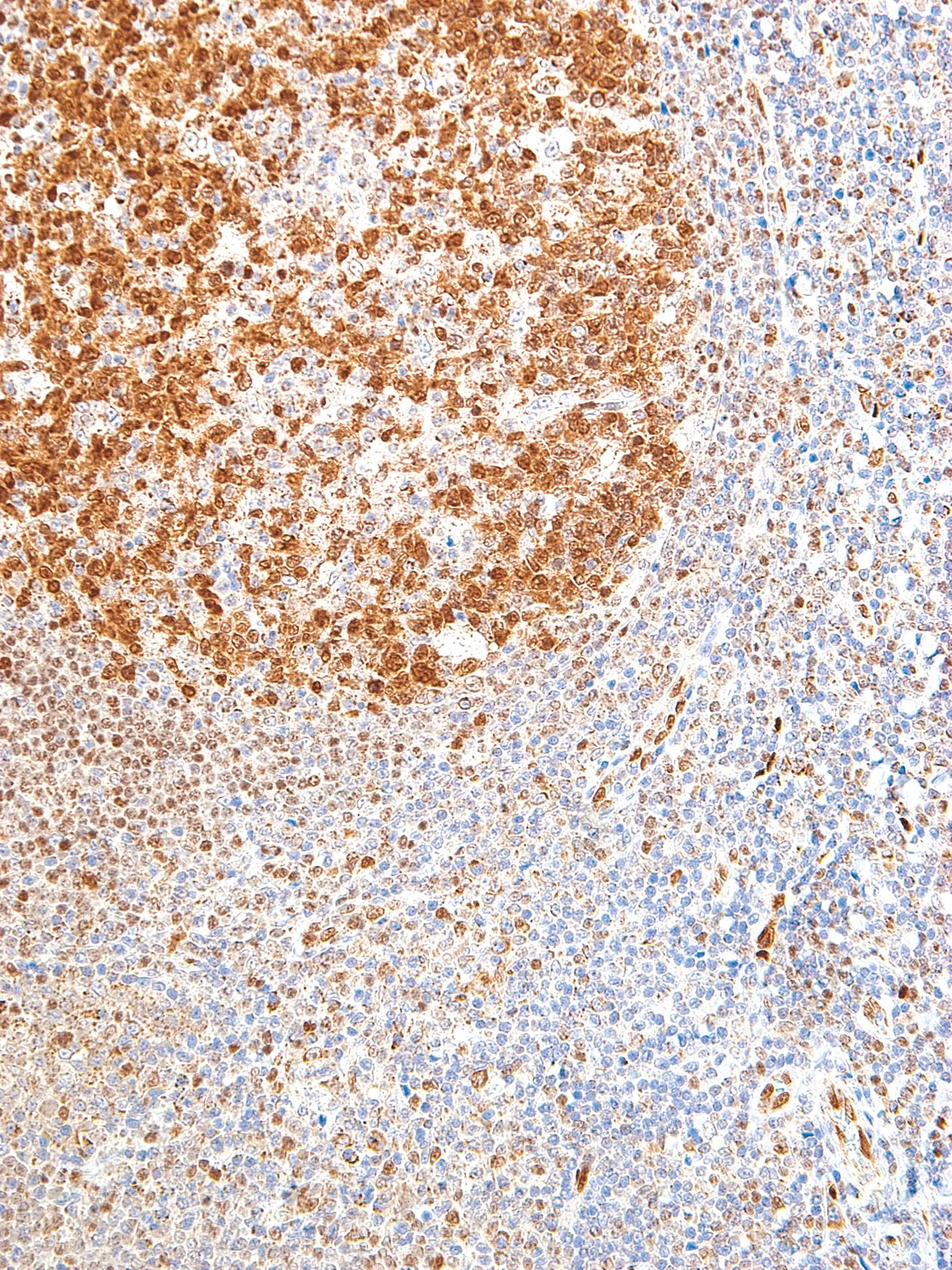
| Clone | IHC615 |
| Source | Mouse Monoclonal |
| Positive Control | Tonsil, Follicular Lymphoma, Diffuse Large B-Cell Lymphoma |
| Dilution Range | 1:200 |

| Clone | IHC615 |
| Source | Mouse Monoclonal |
| Positive Control | Tonsil, Follicular Lymphoma, Diffuse Large B-Cell Lymphoma |
| Dilution Range | 1:200 |


Cell Culture Flask T225
Cell Culture Flask surface is very smooth based on precise molding technology and it gives clear view when examined with microscope.
Volume: 25mL75mL175mL225mL
PRODUCT FEATURES
For adherent cell culture: Initial adherence and proliferative property of cells via hydrophilic surface treatment.
For suspension cell culture: The surface is resistant to cell adherence, which minimizes damage or loss of cell.

Cell Culture Flask surface is very smooth based on precise molding technology and it gives clear view when examined with microscope.
Volume: 25mL75mL175mL225mL
