
Description
Specifications
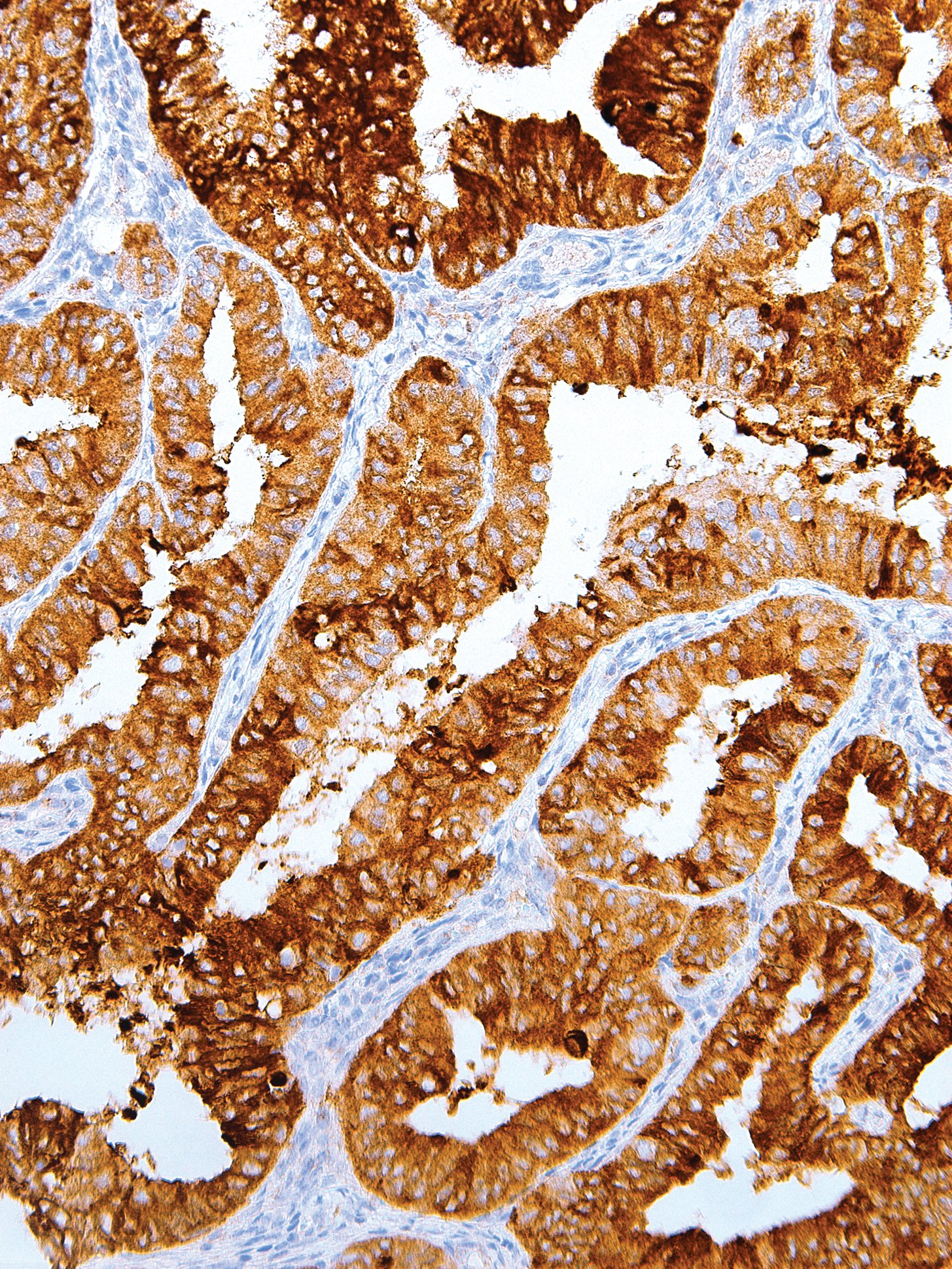
| Clone | IHC623 |
| Source | Mouse Monoclonal |
| Positive Control | Breast, Colon, Associated Adenocarcinomas |
| Dilution Range | 1:200 |

| Clone | IHC623 |
| Source | Mouse Monoclonal |
| Positive Control | Breast, Colon, Associated Adenocarcinomas |
| Dilution Range | 1:200 |

Nucleic acid testing (NAT) is the method of choice for detection and quantification of a wide range of micro organisms. Primerdesign manufactures and supplies high quality quantitative real-time PCR kits for the detection and simultaneous quantification of numerous significant pathogens . A copy number standard curve is provided for quantification and an the internal extraction template (DNA or RNA), controls for the quality of the nucleic acid extraction and eliminates false negative results.
The kit is designed with the broadest possible detection profile to ensure that all clinically relevant strains and subtypes are detected*. Target sequences are selected by working with data from key opinion leaders in the field. Multiple sequence alignments and unprecedented real-time PCR expertise in design and validation ensure the best possible kit.
Details of the target and priming specificity are included in the individual handbooks above.
Packaged, optimised and ready to use. Expect Better Data.
Exceptional value for money
Rapid detection of all clinically relevant subtypes
Positive copy number standard curve for quantification
Highly specific detection profile
High priming efficiency
Broad dynamic detection range (>6 logs)
Sensitive to < 100 copies of target
Accurate controls to confirm findings


This kit provides a single tube to screen for the presence of high-risk HPV types, HPV16, HPV18, HPV31, HPV33, HPV35, HPV39, HPV45, HPV51, HPV52, HPV56, HPV58, HPV59, HPV66 and HPV68. The multiplex test is detected in two fluorescent channels differentiating between HPV16 / HPV18 which both produce a VIC channel signal and all others which produce a signal in the FAM channel. HPV16 and HPV18 account for 70% of positive findings in clinical practice so it is helpful to know if either of these are present. All other high risk genotypes together make up the remaining 30% of clinical positives and are grouped together into the FAM channel. In this configuration, the kit gives a partial genotyping result and some additional information on which high risk strains are present.
Exceptional value for money
Rapid detection of all clinically relevant subtypes
Highly specific detection profile
High priming efficiency
Broad dynamic detection range (>6 logs)
Sensitive to < 100 copies of target
Accurate controls to confirm findings