
Description
Specifications
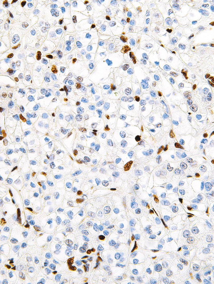
| Clone | IHC672 |
| Source | Mouse Monoclonal |
| Positive Control | Testes, Melanoma, Xp11.2 Translocation Renal Cell Carcinoma |
| Dilution Range | 1:200 |

| Clone | IHC672 |
| Source | Mouse Monoclonal |
| Positive Control | Testes, Melanoma, Xp11.2 Translocation Renal Cell Carcinoma |
| Dilution Range | 1:200 |

Description
The PM5000 ExcelBand™ 3-color Pre-Stained Protein Ladder Regular Range is a ready-to-use three-color protein standard with 13 pre-stained proteins covering a wide range of molecular weights from 10 to 180 kDa in Tris-Glycine Buffer (9 to 170 kDa in Bis-Tris (MOPS) buffer and 10 to 170 kDa Bis-Tris (MES) buffer). Proteins are covalently coupled with different chromophores for easy identification of bands, with three reference proteins carrying enhanced intensity corresponding to a blue band at 20 kDa, green at 40 kDa, and red at 75 kDa, respectively, as separated on SDS-PAGE (Tris-Glycine buffer). The PM5000 ExcelBand™ 3-color Pre-Stained Protein Ladder Regular Range is designed for monitoring protein separation during SDS-polyacrylamide gel electrophoresis, verification of Western transfer efficiency on membranes (PVDF, nylon, or nitrocellulose) and for approximating the size of proteins.
Features
Contents
Approximately 0.1~0.4 mg/ml of each protein in the buffer (20 mM Tris-phosphate (pH 7.5 at 25°C), 2% SDS, 0.2 mM DTT, 3.6 M urea, and 15% (v/v) glycerol).
Quality Control
Under suggested conditions, PM5000 ExcelBand™ 3-color Pre-Stained Protein Ladder Regular Range resolves 13 major bands in SDS-PAGE (Tris-Glycine, MOPS, and MES buffer) and after Western blotting to nitrocellulose membrane.
Storage
4°C for 3 months
-20°C for long term storage
The PM5000 ExcelBand™ 3-color Pre-Stained Protein Ladder Regular Range is a ready-to-use three-color protein standard with 13 pre-stained proteins covering a wide range of molecular weights from 10 to 180 kDa in Tris-Glycine Buffer (9 to 170 kDa in Bis-Tris (MOPS) buffer and 10 to 170 kDa Bis-Tris (MES) buffer). Proteins are covalently coupled with different chromophores for easy identification of bands, with three reference proteins carrying enhanced intensity corresponding to a blue band at 20 kDa, green at 40 kDa, and red at 75 kDa, respectively, as separated on SDS-PAGE (Tris-Glycine buffer). The PM5000 ExcelBand™ 3-color Pre-Stained Protein Ladder Regular Range is designed for monitoring protein separation during SDS-polyacrylamide gel electrophoresis, verification of Western transfer efficiency on membranes (PVDF, nylon, or nitrocellulose) and for approximating the size of proteins.

Opentrons 4-in-1 Tube Rack set includes 2 base stands and 4 tube holder tops: 1.5ml, 2ml, 15ml + 50ml, 15ml, and 50ml. The tube holder tops snap securely on the base stand, which fits directly on the deck. The Tube Rack Set is not autoclavable.
Opentrons 4-in-1 Tube Rack set includes 2 base stands and 4 tube holder tops: 1.5ml, 2ml, 15ml + 50ml, 15ml, and 50ml. The tube holder tops snap securely on the base stand, which fits directly on the deck. The Tube Rack Set is not autoclavable.

African Swine Fever Virus (ASFV) is a widespread disease which infects members of the pig family(Suidae). Anumberoftick species are believed to be the vector for the disease,as well as being transmitted by raw pork and pig excrement [1]. After firstly being identified in Kenya in 1921, ASFV became endemic in sub-Saharan Africa, with regular outbreaks being reported across Europe, Asia and South America throughout the century [2]. More recently the virus was introduced in Georgia and spread throughout the region, as well as mass outbreaks occurring in China in 2018 [3].
ASFVistheonlymemberoftheAsfaridaefamily.ItisalargeenvelopeddoublestrandedDNA virus of icosahedral morphology with an average diameter of 200nm and isolates contain genomes between 170-190Kbp encoding for up to 167 open reading frames [2]. The morphology of ASFV consist of several concentric domains. An inner core contains the nucleoid coated with a thick protein layered core shell, which is surrounded by an inner lipid envelope , all of which is encompassed by the capsid [2]. ASFV begins its replication cycle in the nucleus of infected cells before moving to the cytoplasm where the majority of the replication takes place [2]. Gene transcription is highly regulated, with distinct classes of mRNA identified to accumulate at early, intermediate and late transcripts of the virus [2]. The disease induces acute haemorrhagic disease within its hosts, causing high fevers and skin haemorrhages, with death often occurring within ten days of clinical symptoms appearing [4].
References: 1: The Centre for Food Security and Public Health (2015), African Swine Fever. 2: Galindo, I. and Alonso, C., 2017. African swine fever virus: a review. Viruses, 9(5), p.103. 3: Zhou, X., Li, N., Luo, Y., Liu, Y., Miao, F., Chen, T., Zhang, S., Cao, P., Li, X., Tian, K. and Qiu, H.J., 2018. Emergence of African swine fever in China, 2018. Transboundary and emerging diseases, 65(6), pp.1482-1484. 4: Gallardo, C., Ademun, A.R., Nieto, R., Nantima, N., Arias, M., Martín, E., Pelayo, V. and Bishop, R.P., 2011. Genotyping of African swine fever virus (ASFV) isolates associated with disease outbreaks in Uganda in 2007. African Journal of biotechnology, 10(17), pp.3488-3497.
Exceptional value for money
Rapid detection of all clinically relevant subtypes
Positive copy number standard curve for quantification
Highly specific detection profile
High priming efficiency
Broad dynamic detection range (>6 logs)
Sensitive to < 100 copies of target
Accurate controls to confirm findings