
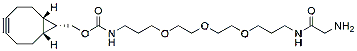
Gly-PEG3-endo-BCN is a click chemistry reagent. The hydrophilic PEG spacer increases solubility in aqueous media. Reagent grade, for research purpose. Please contact us for GMP-grade inquiries.


Gly-PEG3-endo-BCN is a click chemistry reagent. The hydrophilic PEG spacer increases solubility in aqueous media. Reagent grade, for research purpose. Please contact us for GMP-grade inquiries.

K-CellG5-4V
SKU: 700004272
| Content: | (K-CellG5-4V) 120 / 240 assays (manual) / 480 assays (auto-analyser) or (K-CellG5-2V) 60 / 120 assays (manual) / 240 assays (auto-analyser) |
| Shipping Temperature: | Ambient |
| Storage Temperature: | Short term stability: 2-8oC, Long term stability: See individual component labels |
| Stability: | > 2 years under recommended storage conditions |
| Analyte: | endo-Cellulase |
| Assay Format: | Spectrophotometer, Auto-analyser |
| Detection Method: | Absorbance |
| Wavelength (nm): | 400 |
| Signal Response: | Increase |
| Limit of Detection: | 1.2 x 10⁻³ U/mL |
| Reproducibility (%): | ~ 3% |
| Total Assay Time: | 10 min |
| Application examples: | Fermentation broths, industrial enzyme preparations and biofuels research. |
| Method recognition: | Novel method |
The K-CellG5-2V pack size has been discontinued (read more).
Cellulase Activity Assay Kit.
The CellG5 assay reagent for the measurement of endo-cellulase (endo-1,4-β-glucanase) contains two components; 1) 4,6-O-(3-Ketobutylidene)-4-nitrophenyl-β-D-cellopentaoside (BPNPG5) and 2) thermostable β-glucosidase. The ketone blocking group prevents any hydrolytic action by the β-glucosidase on BPNPG5. Incubation with an endo-cellulase generates a non-blocked colourimetric oligosaccharide that is rapidly hydrolysed by the ancillary β-glucosidase. The rate of formation of 4-nitrophenol is therefore directly related to the hydrolysis of BPNPG5 by the endo-cellulase. The reaction is terminated and the phenolate colour is developed on addition of Tris buffer solution (pH 9.0).
The CellG5 assay represents a huge step forward in the methodology for the measurement of cellulase that traditionally relied on substrates such as CM-cellulose, Avicel, cellooligosaccharides, filter paper or dyed polysaccharides including CMC Congo red or cellulose azure.
View Cellulase Activity Assay Protocol.
View our complete list of assay kits for enzyme activities.

The CellG5 assay reagent for the measurement of endo-cellulase (endo-1,4-β-glucanase) contains two components; 1) 4,6-O-(3-Ketobutylidene)-4-nitrophenyl-β-D-cellopentaoside (BPNPG5) and 2) thermostable β-glucosidase. The ketone blocking group prevents any hydrolytic action by the β-glucosidase on BPNPG5. Incubation with an endo-cellulase generates a non-blocked colourimetric oligosaccharide that is rapidly hydrolysed by the ancillary β-glucosidase. The rate of formation of 4-nitrophenol is therefore directly related to the hydrolysis of BPNPG5 by the endo-cellulase. The reaction is terminated and the phenolate colour is developed on addition of Tris buffer solution (pH 9.0).

DBCO-NHCO-PEG13-acid is an analog of DBCO-Acid with a hydrophilic PEG spacer arm, which improves water solubility. This reagent is a non-activated building block with enhanced solubility in aqueous media used to derivatize amine-containing molecule through a stable amide bond. DBCO is commonly used for copper-free Click Chemistry reactions. Reagent grade, for research purpose. Please contact us for GMP-grade inquiries.

DBCO-NHCO-PEG13-acid is an analog of DBCO-Acid with a hydrophilic PEG spacer arm, which improves water solubility. This reagent is a non-activated building block with enhanced solubility in aqueous media used to derivatize amine-containing molecule through a stable amide bond. DBCO is commonly used for copper-free Click Chemistry reactions. Reagent grade, for research purpose. Please contact us for GMP-grade inquiries.

This kit enables the rapid purification of amplified DNA products from PCR mixes. It is able to effectively remove PCR by-products including primers, dimers, enzymes, unincorporated nucleotides and mineral oil from the desired PCR product. The purified PCR products are fully compatible with restriction enzyme digestion, ligation into vectors, labeling, sequencing and more. This kit can also be used as an alternative to organic extraction and ethanol precipitation to clean up various enzymatic reactions.
The kit is also available in a 96-well format for high-throughput PCR purification. Purification with the 96-well plate can be performed using either a vacuum manifold or centrifugation.
Figure 1 / 2
Click for expanded view
| Kit Specifications – Spin Column | |
| Column Binding Capacity | 10 μg |
| Size of DNA Purified | 100 – 15,000 bp |
| Average Recovery | > 90% |
| Average Primer Removal | > 90% |
| Minimum Elution Volume | 30 μL |
| Time to Complete 10 Purifications | 15 minutes |
Storage Conditions and Product Stability
All solutions should be kept tightly sealed and stored at room temperature. This kit is stable for 1 year after the date of shipment.
| Component | Cat. 14400 (50 preps) | Cat. 45700 (250 preps) | Cat. 24800 (192 preps) |
|---|---|---|---|
| Binding Buffer C | 30 mL | 5 x 30 mL | 3 x 30 mL |
| Wash Solution A | 12 mL | 2 x 20 mL | 2 x 38 mL |
| Elution Buffer B | 8 mL | 2 x 30 mL | 2 x 15 mL |
| Spin Columns | 50 | 250 | – |
| Collection Tubes | 50 | 250 | – |
| 96-Well Plate | – | – | 2 |
| Adhesive Tape | – | – | 4 |
| 96-Well Collection Plate | – | – | 2 |
| Elution Tubes (1.7 mL) | 50 | 250 | – |
| 96-Well Elution Plate | – | – | 2 |
| Product Insert | 1 | 1 | 2 |