Usages: For isolating , cultivating mold and yeast .
Principle: Potato flour leaching contribute various mold growth; glucose to provide energy; agar as medium coagulant; chloramphenicol inhibit the growth of bacteria.
Formulation(per liter): Infusion from potatoes 200g Glucose 20g Agar 15g Final pH 5.6 ± 0.2
How to use: 1.Suspend 40g in 1L of distilled water , stirring heated to boiling to completely dissolve ,autoclave at 121 for 15 minutes. 2.Diluted and treated samples.
Storage: Keep container tightly closed, store in a cool, dry place, away from bright light. Storage period of 3 years.
Other Products
TD4 serofuge
Product Info
Document
Product Info
Cell Washer/Serofuge Centrifuge
Features
1. Microprocessor control, less noisily, it is widely used to qualitative analysis of blood serum, plasma and urea in the fields of hospital, laboratory and biochemistry.
2. Brushless motor, free maintenance, no powder pollution, quick in speed up and down.
3. Special program, HLA and SERO separately.
5. Micro computer control system, LED display the RCF,time and speed.
6.. Electric lid lock, compact design, super speed and imbalance protection.
TD4 Technical Parameter:
Max. Speed
4000rpm
Max. RCF
2000
Max. Capacity
12×7
Time Range
0~99min
RPM/RCF Convert
Yes
Noise (dB)
≤ 55
Temperature
Normal
Acc/Dcc
10 Kinds
Speed Accuracy
±20r/min
Temperature Accuracy
/
Voltage(V/Hz)
AC 220V/110V 50HZ/60HZ
Size (W x D x Hmm)
485×320×255mm
Net Weight(Kg)
24KG
Matched Rotors for TD4
Order No.
Rotor
Max Speed (rpm)
Max Volume(ml)
Max. RCF(×g)
NO31501
SERO Rotor
3000
12×7/5ml(10-13*60-100mm)
2000
NO31502
HLA Rotor
4000
12×2/1.5/0.5ml
1000
Rotor Name
Max RCF(×g)
Use
HLA rotor for Lymphocyte washing
2000
Lymphocyte separation incubated cell separation
1000
Hematoblast extenterate(thrombin disposal)
1000
Lymphocyte washing
SERO rotor for erythrocyte washing
500
Blood type determination, the image of the hematocyte
1000
The test of intersectant aptness
1000
Hematocyte washing, the distillation of the serum and plasma
Document
TD4 cell washer centrifuge it is widely used to qualitative analysis of blood serum, plasma and urea in the fields of hospital, laboratory and biochemistry.
Rapid spin column purification of genomic DNA from viable yeast cells, fungal spores or mycelium, and bacteria including Gram-positive
Bead tubes (provided) allow for effective mechanical homogenization
Purified DNA is of high quality and integrity and compatible with any sensitive downstream applications such as PCR, qPCR, RFLP and more
These kits provide fast and reliable procedures for the purification of genomic DNA from viable yeast cells, fungal spores or mycelium, and bacteria including Gram-positive. Genomic DNA is efficiently extracted from the cells by a combination of heat treatment, detergents and Bead Tubes (provided). An optional lyticase treatment allows for improved DNA yields with certain fungal and yeast species. Recovered genomic DNA is of excellent yield and purity for any downstream application including PCR, qPCR, Restriction Fragment Length Polymorphism (RFLP), Amplified Fragment Length Polymorphism (AFLP), sequencing, SNP analysis and more.
Fungi/Yeast Genomic DNA Isolation Kit (Spin Column)
This kit provides rapid spin column purification of genomic DNA from viable yeast cells, fungal spores or mycelium, and bacteria including Gram-positive. Preparation time for a single sample is less then 30 minutes, and each kit contains sufficient materials for 50 preparations.
Fungi/Yeast Genomic DNA Isolation 96-Well Kit (HT)
Norgen’s Fungi/Yeast Genomic DNA Isolation 96-Well Kit provides a fast, reliable and simple procedure for high throughput isolation of DNA from viable yeast cells, fungal spores or mycelium and Gram-positive bacteria. The purification could be performed on either a vacuum manifold or using centrifugation. Complete 96 purifications in 40 minutes.
Maximum Amount of Starting Material: Fungi (Wet weight) Yeast or Gram-positive bacterial culture
50 mg 0.5 mL – 1 mL
Time to Complete 96 Purifications
40 minutes
Storage Conditions and Product Stability All solutions should be kept tightly sealed and stored at room temperature. These reagents should remain stable for at least 2 years in their unopened containers.
Plasmid Purification Magnetic Beads (RNA Depletion)
Product Info
Document
Product Info
Plasmid Purification Magnetic Beads (RNA Depletion)
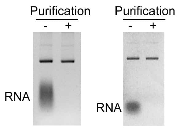
Plasmid isolation from bacterial cultures is one of the most popular techniques in biomedical research and pharmaceutical industries. However, it is common that the isolated plasmid DNA is usually contaminated with varied degrees of host RNA. Plasmid purification is necessary to reduce the impact on downstream applications by removing RNA contamination.
We have developed a simple reagent to completely remove RNA contamination in the isolated plasmid samples using Solid Phase Reversible Immobilization (SPRI) beads. SPRI beads consist of paramagnetic particles coated with carboxyl groups that reversibly bind DNA. Our Plasmid Purification Magnetic Beads (RNA Depletion) combines BioDynami’s proprietary chemistries with the reversible DNA-binding properties of SPRI magnetic beads. The reagent removes RNA and recovers the plasmid in the same step. Moreover, unwanted components such as salts, dNTPs, proteins, enzymes, and other impurities can also be removed simultaneously.
Plasmid can be used for downstream applications such as enzymatic digestion, transformation, transfection and molecular cloning etc. The beads can be an effective and inexpensive reagent for bacterial RNA depletion for routine plasmid purification.
Features
Effective depletion of bacterial RNA by RNase
High recovery rate of plasmid DNA by magnetic beads
Removal of unwanted components and impurities
Simple and fast beads-based protocol
Document
Plasmid isolation from bacterial cultures is one of the most popular techniques in biomedical research and pharmaceutical industries. However, it is common that the isolated plasmid DNA is usually contaminated with varied degrees of host RNA. Plasmid purification is necessary to reduce the impact on downstream applications by removing RNA contamination.


 Matched Rotors for TD4