Usages: For the differentiation of Gram-negative bacteria on the basis of citrate utilization.
Principle: Magnesium ions in various metabolic cofactors; ammonium dihydrogen phosphate to provide nitrogen; dipotassium hydrogen phosphate is the buffer; sodium citrate as a carbon source; agar as medium coagulant.
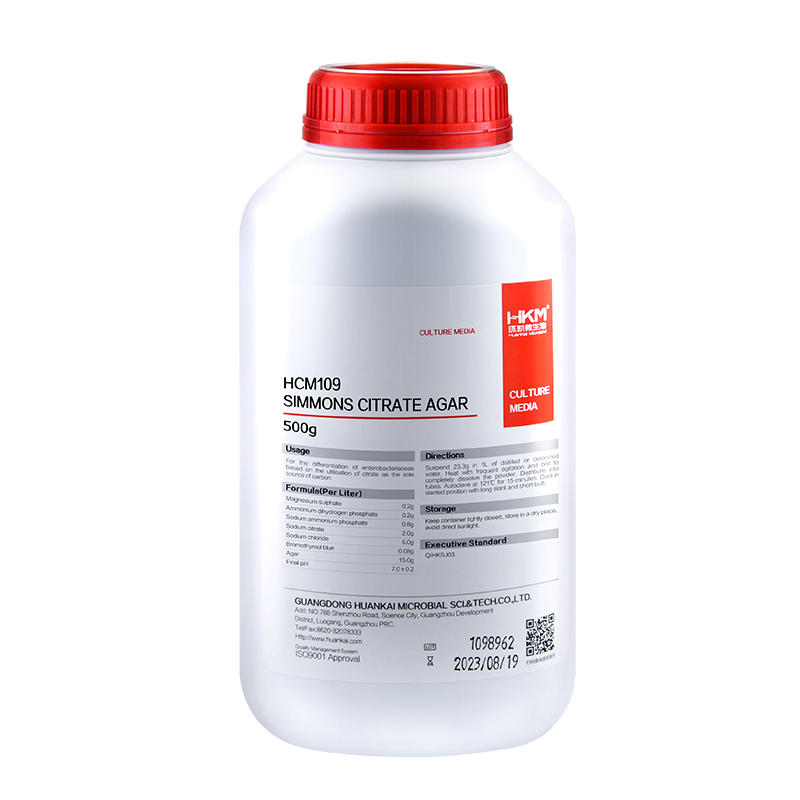
Formulation(per liter): Sodium chloride 5g Magnesium sulfate 0.2g Ammonium dihydrogen phosphate 0.2g Sodium ammonium phosphate 0.8g Sodium citrate 2g Agar 15g Bromothymol blue 0.08g Final pH 7.0 ± 0.2
How to use: 1.Suspend 23.3g in 1 L of distilled water , stirring heated to boiling until completely dissolved, dispensing flask, 121 autoclave for 15min. 2.Diluted and treated samples.
Storage: Keep container tightly closed, store in a cool, dry place, away from bright light. Storage period of 3 years.
Other Products
P1811 MagPure Plasmid Mini Kit
Product Info
Document
Product Info
Introduction
The MagPure Plasmid purification system uses the paramagnetic bead technology for high-throughput preparation of high-copy or low-copy plasmid DNA from E. coli cells. This kit also can be used with fosmid and BAC vector-based constructs. The system uses alkaline lysis followed by a MagPure purification to differentially bind plasmid DNA to paramagnetic beads. While the DNA is bound to the beads, contaminants can be rinsed away using a simple washing procedure. Because MagPure uses magnetic separation technology, the protocol does not require vacuum filtration. This makes kit extremely amenable to automation. Plasmid DNA purified with this system is most commonly used in Sanger Sequencing and PCR amplification.
Details
Specifications
Features
Specifications
Main Functions
Isolation up to 20µg plasmid DNA from 1-3ml bacterial culture
Applications
Enzyme digestion, sequencing, PCR and labeling, etc.
Purification technology
Magnetic beads technology
Process method
Manual or automatic
Sample type
Conventional plasmid, plasmid≤30KB
Sample amount
1-3ml
Elution volume
≥50μl
Principle
This product is based on the purification method of high binding magnetic particles. The sample is lysed and digested under the action of lysate and Lysozyme. DNA is released into the lysate. After adding magnetic particles and binding solution, DNA will be adsorbed on the surface of magnetic particles, and impurities such as proteins will be removed without adsorption.The adsorbed particles were washed with washing solution to remove proteins and impurities, washed with ethanol to remove salts, and finally DNA was eluted by Elution Buffer.
Advantages
1. Suitable for extracting plasmids from 1-5ml or <3ml YT medium. 2. The same amount of buffer 1, 2, and 3 avoids errors caused by adjusting the pipette, making it convenient to use in conjunction with automated workstations. 3. Containing buffer 1 for washing, reducing the problem of false high production. 4. The purified plasmid can be directly used for sequencing, enzyme digestion, PCR, and other applications.
Kit Contents
Contents
P181102
P181103
P181104
Purification Times
100 Preps
500 Preps
5000 Preps
RNase A
10 mg
50 mg
2 x 250 mg
Buffer P1
30 ml
150 ml
2 x 750 ml
Buffer P2
30 ml
150 ml
2 x 750 ml
Buffer N3
30 ml
150 ml
2 x 750 ml
Buffer PW1
35 ml
180 ml
2 x 900 ml
MagPure Particle NB*
2.2 ml
11 ml
2 x 60 ml
Storage and Stability
RNase A and MagPure Particle NB should be stored at 2-8°C upon arrival. However, short-term storage (up to 24 weeks) at room temperature (15-25°C) does not affect its performance. The remaining kit components can be stored dry at room temperature (15-25°C) and are stable for at least 18 months under these conditions. After addition of RNase A, Buffer P1 is stable for 6 months when stored at 2-8°C.
Experiment Data
Document
The MagPure Plasmid purification system uses the paramagnetic bead technology for high-throughput preparation of high-copy or low-copy plasmid DNA from E. coli cells. This kit also can be used with fosmid and BAC vector-based constructs. The system uses alkaline lysis followed by a MagPure purification to differentially bind plasmid DNA to paramagnetic beads. While the DNA is bound to the beads, contaminants can be rinsed away using a simple washing procedure. Because MagPure uses magnetic separation technology, the protocol does not require vacuum filtration. This makes kit extremely amenable to automation. Plasmid DNA purified with this system is most commonly used in Sanger Sequencing and PCR amplification.
Introducing the Fastin Assay Kit: Your Straightforward Solution for Elastin Quantification! Our user-friendly kit utilizes a dye-based method to measure elastin from in-vivo and in-vitro sources. It can be used to quantify various elastin forms, spanning from immature tropoelastin to mature, ‘insoluble’ elastin fibers.
Colorimetric Detection (513nm) (Endpoint)
Understanding Elastin: The Key to Tissue Flexibility
Tissues like lungs and arteries must maintain the ability to stretch and recoil repeatedly throughout an organism’s life. Elastin, a mature protein, is responsible for this elasticity and is usually present as insoluble fibers within the ECM. During development, these fibers are initially formed from a soluble precursor called tropoelastin.
What is the Fastin Assay?
The Biocolor Fastin assay is a user-friendly, dye-based means of quantifying elastins derived from both in-vivo and in-vitro sources. A variety of elastin forms can be assayed, from immature tropoelastin to mature ‘insoluble’ elastin fibres.
Further information on how the assay works can be found on the ‘Mode of Action‘ tab.
A list of suggested sample types can be found under the ‘Assay Specification‘ tab.
How does the Fastin assay detect Elastin?
The Fastin Dye Reagent contains an elastin-binding synthetic porphyrin, TPPS (5,10,15,20-tetraphenyl- 21H,23H-porphine). This affinity of TPPS for elastin was first observed when used as a ‘vital stain’ on live animals. Most tissues took up the dye initially but only elastin retained the TPPS molecules over time. [Winkelman, J. (1962), Cancer Res. 22, 589-596; Winkelman, J & Spicer, S. (1962), Stain Technol. 37, 303-305].
It has been proposed that the elastin binding of TPPS may be due to the retention of the acidic dye (which contains four charged sulfate groups) by the basic amino acid side chain residues of elastin.
How does the Fastin assay work?
Step 1. Incubation of samples containing soluble elastin with the Fastin Dye Reagent causes an elastin-dye complex to form. This insoluble complex then precipiates.
Step 2. Dye-labelled elastin is then isolated by centrifugation and the unbound dye removed. Elastin-bound dye is then eluted and measured spectrophotometrically.
Step 3. The elastin content of unknown samples can be calculated by comparison against a calibration curve prepared using a standard comprising water-soluble elastin (supplied with the kit).
Assay range
50 – 500µg/ml
Limit of Detection
50µg/ml
Detection Method
Colorimetric Detection (513nm) (Endpoint)
Measurements per kit
110 in total (allows a maximum of 48 samples to be run in duplicate alongside a standard curve).
Suitable Samples
In-vivo: tissues and fluids. Insoluble elastin will first require conversion to water soluble α-elastin using the oxalic acid reagents and extraction protocol supplied with the kit.
In-vitro: Elastin produced by cells during 2D/3D cell culture. NB elastin in conditioned cell media is typically below the detection limit of the kit.
Precautions
This kit is designed for research use only. Not for use in diagnostic procedures. Kit requires access to a centrifuge, heated water bath or block, as well as a spectrophotometer or colorimeter capable of absorbance detection at 513nm. Specific sample preparation protocols may require customer to provide further reagents, consult assay manual for further information.
Fastin elastin kit contents:
1. Dye Reagent (1x110ml)
2. α-elastin Reference Standard (1x5ml, 1.0 mg/ml soluble Bovine elastin)
6. 1.5ml micro-centrifuge tubes for dye-labelling reaction.
7. Assay kit manual
NB: Additional reagents may be required for sample preparation prior to assay. Consult manual or contact us for further details.
Document
Introducing the Fastin Assay Kit: Your Straightforward Solution for Elastin Quantification! Our user-friendly kit utilizes a dye-based method to measure elastin from in-vivo and in-vitro sources. It can be used to quantify various elastin forms, spanning from immature tropoelastin to mature, ‘insoluble’ elastin fibers. Colorimetric Detection (513nm) (Endpoint)
The 16S ribosomal RNA (rRNA) plays a crucial role in bacterial and archaeal ribosomes. This sequence is Highly conserved across bacteria and archaea, it contains variable regions useful for species differentiation and is part of the 30S subunit of prokaryotic ribosomes. 16s rRNA Binds to the Shine-Dalgarno sequence and contributes to the subunit’s structure. Acts as a scaffold for ribosomal proteins.
Because the 16s rRNA sequence is ubiquitous in bacteria and archaea, it can be used to identify a wide diversity of microbes within a single sample and single workflow.
Document
This kit is sufficient for 150 reactions: For characterizing cyanobacteria in environmental samples Use in combination with Attogene Algae DNA isolation kit Universal 16s PCR primers Perfect for Environmental DNA (eDNA) Characterization