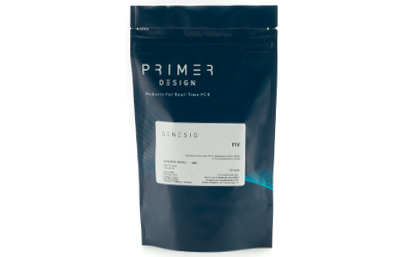
Primer and probe mix (150 reactions) Reverse Transcription, target specific primers (RNA genome viruses only) Copy number standard curve (sufficient for multiple standard curves) Internal extraction control – Read through VIC channel* Endogenous control (150 tests) RNAse/DNAse free water *alternative fluorophores available on request
Nucleic acid testing (NAT) is the method of choice for detection and quantification of a wide range of micro organisms. Primerdesign manufactures and supplies high quality quantitative real-time PCR kits for the detection and simultaneous quantification of numerous significant pathogens . A copy number standard curve is provided for quantification and an the internal extraction template (DNA or RNA), controls for the quality of the nucleic acid extraction and eliminates false negative results.
The kit is designed with the broadest possible detection profile to ensure that all clinically relevant strains and subtypes are detected. Target sequences are selected by working with data from key opinion leaders in the field. Multiple sequence alignments and unprecedented real-time PCR expertise in design and validation ensure the best possible kit. Details of the target and priming specificity are included in the individual handbooks above.
Packaged, optimised and ready to use. Expect Better Data.
Other Products
D6323D MagPure FFPE DNA Kit (High Pure)
Product Info
Document
Product Info
Introduction
This product is suitable for rapid extraction of DNA from FFPE sample. This kit uses two combination methods. High-salt Bind is conducive to remove pigments or polysaccharides from complex FFPE samples, so as to improve the purity of nucleic acid and avoid blocking aligent 2100. Alcohol mediated adsorption is conducive to improve the nucleic acid yield of high-yield samples.
Details
Specifications
Features
Specifications
Main Functions
Isolation high pure total DNA from FFPE using high bind beads
Applications
PCR and viral DNA detection, etc.
Purification technology
Magnetic beads technology
Process method
Manual or automatic
Sample type
Paraffin embedded tissue samples
Sample amount
1-6 slices of 10-20μm
Elution volume
≥30μl
Time per run
≤60 minutes
Principle
This product is based on the purification method of high binding magnetic particles. The sample is lysed and digested under the action of lysate and Protease. DNA is released into the lysate. After adding magnetic particles and binding solution, DNA will be adsorbed on the surface of magnetic particles, and impurities such as proteins will be removed without adsorption. The adsorbed particles were washed with washing solution to remove proteins and impurities, washed with ethanol to remove salts, and finally DNA was eluted by Elution Buffer.
Advantages
High yield – most optimal process, recovery up to 90%
Economy – less than 50% of the price of Qiagen and other imported products
High purity – OD 260/280 : 1.7-1.9, OD 260/230 : 1.5-2.0
Kit Contents
Contents
D632301D
D632302D
Purification Times
48 Preps
96 Preps
MagPure Particles N
1.1 ml
2.5 ml
RNase A
10 mg
20 mg
Proteinase K
24 mg
48 mg
Protease Dissolve Buffer
3 ml
6 ml
Buffer DPS
60 ml
100 ml
Buffer ATL
15 ml
30 ml
Buffer BST1
30 ml
60 ml
Buffer BW1
13 ml
44 ml
Elution Buffer
15 ml
30 ml
Storage and Stability
Proteinase K, RNase A and MagPure Particles N should be stored at 2-8°C upon arrival. However, short-term storage (up to 12 weeks) at room temperature (15-25°C) does not affect their performance. The remaining kit components can be stored at room temperature (15-25°C) and are stable for at least 18 months under these conditions.
Document
This product is suitable for rapid extraction of DNA from FFPE sample. This kit uses two combination methods. High-salt Bind is conducive to remove pigments or polysaccharides from complex FFPE samples, so as to improve the purity of nucleic acid and avoid blocking aligent 2100. Alcohol mediated adsorption is conducive to improve the nucleic acid yield of high-yield samples.
Nucleic acid testing (NAT) is the method of choice for detection and quantification of a wide range of micro organisms. Primerdesign manufactures and supplies high quality quantitative real-time PCR kits for the detection and simultaneous quantification of numerous significant pathogens . A copy number standard curve is provided for quantification and an the internal extraction template (DNA or RNA), controls for the quality of the nucleic acid extraction and eliminates false negative results.
The kit is designed with the broadest possible detection profile to ensure that all clinically relevant strains and subtypes are detected. Target sequences are selected by working with data from key opinion leaders in the field. Multiple sequence alignments and unprecedented real-time PCR expertise in design and validation ensure the best possible kit. Details of the target and priming specificity are included in the individual handbooks above.
Packaged, optimised and ready to use. Expect Better Data.
Document
Primer and probe mix (150 reactions) Reverse Transcription, target specific primers (RNA genome viruses only) Copy number standard curve (sufficient for multiple standard curves) Internal extraction control – Read through VIC channel* Endogenous control (150 tests) RNAse/DNAse free water *alternative fluorophores available on request