20 tests Test for the semi-quantitative evaluation of human Interleukin-6 (IL-6) in serum, plasma, cell culture supernatant, amnitotic fluid or cerebrospinal fluid.
Detail
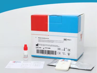
Name of Product
IL-6 / Interleukin-6 Test
Catalog Number
MQL6 1
Short Info
Test for the semi-quantitative evaluation of human Interleukin-6 (IL-6) in serum, plasma, cell culture supernatant, amnitotic fluid or cerebrospinal fluid.
This product is only available within the EU!
Method/Platform
lateral flow
Range/Assay Sensivity
50 to 10 000 pg/ml
Test Principle
IL-6 binds to a first anti IL-6 antibody conjugated to gold particles. The IL-6 loaded gold particles diffuse through the membrane and overflow the test line.
A second monoclonal antibody, specific for IL-6 is coated on the membrane. The gold particles are bound specifically and become visible as a coloured line. Colour intensity is directly proportional to the concentration of IL-6 in the sample. The conjugated specific antibodies printed as a second line on the membrane captures the rest of gold conjugate.
Other Products
Cells and Tissue DNA Isolation Kits
Product Info
Document
Product Info
Overview
Fast, reproducible and easy processing of samples
High yield and high quality genomic DNA with no RNA or protein contamination
DNA ready for any application including PCR, qPCR, genotyping and more
Norgen’s Cells and Tissue DNA Isolation Kits are designed for the rapid preparation of genomic DNA from cultured cells as well as various tissue samples and urine. The purified genomic DNA is fully digestible with all restriction enzymes tested, and is completely compatible with PCR and Southern Blot analysis.
Cells and Tissue DNA Isolation Kit (Spin Column)
Purification is based on spin column chromatography as the separation matrix. Norgen’s columns bind DNA under optimized salt concentrations and release the bound DNA under low salt and slightly alkali conditions. The protocol can be completed in approximately 30 minutes for cells and within 90 minutes for tissues. Each kit contains sufficient materials for 50 preparations.
Cells and Tissue DNA Isolation Micro Kit (Micro)
Optimized for small inputs of cells and tissues, such as Laser-Captured Microdissection (LCM). Purification is based on spin column chromatography as the separation matrix. Norgen’s columns bind DNA under optimized salt concentrations and release the bound DNA under low salt and slightly alkali conditions. Preparation time for a single sample is approximately 60 minutes, and each kit contains sufficient materials for 50 preparations.
Cells and Tissue DNA Isolation Kit (Magnetic Bead System)
Purification is based on the use of magnetic beads that bind DNA under optimized binding conditions. Norgen’s Cells and Tissue DNA Isolation Kit (Magnetic Bead System) allows for the isolation of genomic DNA from various types of animal tissues or cell samples. Preparation for 10 purifications is approximately 40 minutes of hands-on time.
Cells and Tissue DNA Isolation 96-Well Kit (High Throughput Magnetic Bead System)
Purification is based on the use of magnetic beads that bind DNA under optimized binding conditions. Norgen’s Cells and Tissue DNA Isolation 96-Well Kit (Magnetic Bead System) allows for the isolation of genomic DNA from various types of animal tissues or cell samples. The Cells and Tissue DNA Isolation 96-Well Kit (Magnetic Bead System) also can be integrated with a robotic automation system.
8-10 µg (20 mg of animal tissue) 8-12 µg (3 x 10⁶ cells)
Average Purity (OD260/280)
1.8 – 1.9
Time to Complete 10 Purifications
40 minutes hands-on time (Cat. 59100) 50 minutes hands on time (Cat. 62500)
* Average DNA yield will vary depending on the donor
Storage Conditions and Product Stability All solutions should be kept tightly sealed and stored at room temperature (15 – 25°C). This kit is stable for 1 year after the date of shipment.
Amino-Tri-(Propargyl-PEG2-ethoxymethyl)-methane TFA Salt
Product Info
Document
Product Info
Amino-Tri-(Propargyl-PEG2-ethoxymethyl)-methane TFA Salt is a crosslinker consisting of an amino group with three propargyl groups. The amino group is reactive with carboxylic acids, activated NHS esters, carbonyls (ketone, aldehyde) etc. The propargyl groups can form triazole linkage with azide-bearing compounds or biomolecules via copper catalyzed Click Chemistry. Reagent grade, for research purpose. Please contact us for GMP-grade inquiries.
Document
Amino-Tri-(Propargyl-PEG2-ethoxymethyl)-methane TFA Salt is a crosslinker consisting of an amino group with three propargyl groups. The amino group is reactive with carboxylic acids, activated NHS esters, carbonyls (ketone, aldehyde) etc. The propargyl groups can form triazole linkage with azide-bearing compounds or biomolecules via copper catalyzed Click Chemistry. Reagent grade, for research purpose. Please contact us for GMP-grade inquiries.