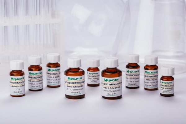
The L-Glutamine/Ammonia (Rapid) test kit is a novel method for the specific, convenient, cost effective and rapid measurement and analysis of L-glutamine and ammonia in culture media/supernatants and other materials.
Detail
K-GLNAM
SKU: 700004295
50 assays of each (manual) / 500 assays of each (microplate)
Content:
50 assays of each (manual) / 500 assays of each (microplate)
Shipping Temperature:
Ambient
Storage Temperature:
Short term stability: 2-8oC, Long term stability: See individual component labels
Stability:
> 2 years under recommended storage conditions
Analyte:
Ammonia, L-Glutamine
Assay Format:
Spectrophotometer, Microplate
Detection Method:
Absorbance
Wavelength (nm):
340
Signal Response:
Decrease
Linear Range:
1 to 40 µg of L-glutamine per assay
Limit of Detection:
0.54 mg/L (L-glutamine), 0.06 mg/L (ammonia)
Reaction Time (min):
~ 10 min
Application examples:
Cell culture media and cultures, dietary supplements, vegetables and other materials (e.g. biological samples, etc.).
Method recognition:
Novel method
This product has been discontinued.
The L-Glutamine/Ammonia (Rapid) test kit is a novel method for the specific, convenient, cost effective and rapid measurement and analysis of L-glutamine and ammonia in culture media/supernatants and other materials.
Note for Content: The number of manual tests per kit can be doubled if all volumes are halved. This can be readily accommodated using the MegaQuantTM Wave Spectrophotometer (D-MQWAVE).
Extended cofactors stability. Dissolved cofactors stable for > 1 year at 4oC.
Very rapid reaction due to use of high activity glutaminase and uninhibited glutamate dehydrogense
All enzymes supplied as stabilised suspensions
Only enzymatic kit available
Very cost effective
All reagents stable for > 2 years after preparation
Mega-Calc™ software tool is available from our website for hassle-free raw data processing
Standard included
Other Products
BKV TaqMan PCR Detection Kits
Product Info
Document
Product Info
Overview
Detection kits for the BKV
Available in TaqMan format for analysis
The BK virus is a member of the polyomavirus family. BK viral infections are typically asymptomatic in healthy individuals, however very mild symptoms may appear including mild respiratory infections and fever. Once an individual has been infected the virus disseminates to the kidneys and the urinary tract where it remains for the lifetime of the individual. Infections with BK virus in immunocompromised or immunosupressed patients are much more severe and may involve renal dysfunction. In fact, in kidney transplant patients the immunosupressive drugs required for the transplant may allow the virus to replicate within the graft, resulting in a disease called BK virus nephropathy (BKVN). It is thought that 1-10% of renal transplant patients progress to BK virus nephropathy (BKVN) and up to 80% of these patients are reported to have lost their grafts. The onset of nephritis can occur as early as several days post-transplant to as late as 5 years. The mode of transmission of the virus is not clear, however it has been suggested that BKV may be transmitted through respiratory fluids or urine, since infected individuals periodically excrete virus in the urine. This virus can be diagnosed by BKV blood & urine testing, in addition to carrying out a biopsy in the kidneys. PCR techniques are now widely used to identify the virus.
BKV TaqMan PCR Kit, 100 reactions
Ready to use format, including Master Mix for the target and PCR control to monitor for PCR inhibition and validate the quality
Specific Primer and Probe mix for the pathogen/virus/viroid of interest
Primer and Probe mix
Positive and negative control to confirm the integrity of the kit reagents
BKV TaqMan PCR Probe/Primer Set and Controls, 100 reactions
Specific Primer/Probe mix and Positive Control for the pathogen/virus/viroid of interest
Nuclease-free water
Can be used together with Norgen’s PCR Master Mix (#28007) or customer supplied master mix
For research use only and NOT intended for in vitro diagnostics.
Storage Conditions and Product Stability All kit components can be stored for 2 years after the date of production without showing any reduction in performance.
All kit components should be stored at -20°C upon arrival.
Our Blyscan™ Glycosaminoglycan Kit has been a ‘go-to’ Solution for reliable sGAG and Proteoglycan Analysis for many years! Blyscan utilises a dye-binding approach to quantitatively measure sulfated glycosaminoglycans (sGAG) and proteoglycans in cells, tissues and fluids from a wide range of in-vivo and in-vitro sources.
Colorimetric Detection (656nm) (Endpoint)
Understanding Glycosaminoglycans (GAGs) and Proteoglycans
Glycosaminoglycans (GAGs) are a type of negatively charged polysaccharide that play crucial roles in various biological processes. They are composed of repeated disaccharide units, typically of N-acetylated or N-sulfated hexosamine paired with a uronic acid (GlcA or IdoA) or galactose. Sulfate groups can also be added to give sulfated GAGs an overall negative charge that influences cell interactions and also enable binding by our Blyscan dye reagent.
Common examples of GAGs include Chondroitin Sulfate, Dermatan Sulfate, Heparin, Heparan Sulfate, and Keratan Sulfate. Note that Hyaluronic Acid is a non-sulfated GAG and cannot be detected by the Blyscan assay. If you need to measure hyaluronic acid instead, we recommend using our Purple-Jelley kit!
The Role of Glycosaminoglycans in Tissues
GAGs and proteoglycans have essential functions in tissues and organisms, providing biophysical support through scaffolding and maintaining cartilage hydration. They also play a vital role in biochemical processes such as cell adhesion and signalling.
What is the origin of the Blyscan assay name?
Blyscan is an Old English word meaning ‘to shine’ and from which the word ‘blush’, (blushing), may have been derived. This was an appropriate choice as the Blyscan Assay contains a blue dye which ‘blushes’ bright pink when it binds to sulphated glycosaminoglycans!
How does the Blyscan assay work?
Step 1. Blyscan dye reagent contains DMMB dye in an optimised buffer. Addition of Dye reagent to samples containing sGAG results in the formation of a dye/sGAG complex due to a charge interaction between dye and GAG sulfate groups.
Step 2. Over a 30 minute incubation Dye-labelled sGAGs precipitate out of solution and are collected by centrifugation. Following removal of unbound dye, the remaining bound dye is released from the complex by addition of dye dissociation reagent. Released dye is quantified spectrophotometrically.
Step 3. The sGAG content of unknown samples may be quantified by comparison against a calibration curve prepared using a standard of purified Chondroitin-4-sulfate supplied with the kit.
A list of suggested sample types can be found under the ‘Assay Specification‘ tab.
The Blyscan Dye reagent is formulated to miminise binding to other charged sample components such as nucleic acids, a problem with some older dye-based sGAG assays.
Assay range
2.5 – 50µg/ml
Limit of Detection
2.5µg/ml
Detection Method
Colorimetric Detection (656nm) (Endpoint)
Measurements per kit
110 in total (allows a maximum of 48 samples to be run in duplicate alongside a standard curve).
In-vivo: Liquid samples, including fluids such as urine, amniotic or synovial fluid.
In-vitro: Solid samples, such as deposited ECM on 2D/3D culture surfaces.by enzymatic treatment
In-vivo: Liquid samples, Culture media during 2D/3D cell culture.
The assay requires that sulfated polysaccahrides or sGAGs are in a soluble form. A preliminary enzymatic extraction step is required for solid samples (enzyme not supplied with kit).
The assay is not suitable for use with samples containing alginates or that comprise degraded sulfated disaccharide fragments.
Precautions
This kit is designed for research use only. Not for use in diagnostic procedures. Kit requires access to a centrifuge, as well as a spectrophotometer/colorimeter capable of absorbance detection at 656nm. Specific sample preparation protocols may require customer to provide further reagents, consult assay manual for further information.
Blyscan sGAG kit contents:
1. Blyscan Dye Reagent (1x110ml)
2.sGAG Reference Standard (1x5ml, 100µg/ml Bovine tracheal chondroitin 4-sulfate)
3. Dissociation Reagent (1x110ml)
4. Sodium Nitrite (1x15ml)
5. Acetic Acid (1x15ml)
6. Ammonium Sulfamate (1x15ml)
7. 1.5ml micro-centrifuge tubes for dye-labelling reaction.
8. Assay kit manual
NB: Additional reagents may be required for sample preparation prior to assay. Consult manual or contact us for further details.
Document
Our Blyscan™ Glycosaminoglycan Kit has been a ‘go-to’ Solution for reliable sGAG and Proteoglycan Analysis for many years! Blyscan utilises a dye-binding approach to quantitatively measure sulfated glycosaminoglycans (sGAG) and proteoglycans in cells, tissues and fluids from a wide range of in-vivo and in-vitro sources. Colorimetric Detection (656nm) (Endpoin
The series of DNA Size Selection Kits (Magnetic Beads) were developed for DNA size selection using magnetic beads. A total of 11 kits are available, with different selection ranges spanning from 50 bp to over 10 kb. The kits provide a simple and quick approach for the enrichment of a specific range of DNA fragments. The kit workflow allows double-sided or single-sided size selection for specific size cutoffs.
Gel images of different ranges of size selection. Sheared human genomic DNA was used as input.
.
DNA size selection is a selective capture of DNA fragments of a specific range of size for next-generation sequencing (NGS) library preparations, PCR, ChIP assay, DNA ligations, endonuclease digestions, adapter removal, and other genomics and molecular biology applications. DNA size selection is preferred after NGS library prep in most of the cases. The NGS library preparation is related to the quality of the sequencing data. Precise NGS library size selection can increase sequencing efficiency, improve data quality, and reduce costs.
There are two types of sequencing technologies: short-read sequencing and long-read sequencing. Short-read sequencing uses DNA libraries that contain small insert DNA fragments of similar sizes, usually several hundred base pairs. The sequencing efficiency can be improved if the DNA size selection is in the right range. Cat.# 20104S and 20104L are the best kits for NGS library size selection of illumina paired-end 100 (PE100) sequencing with 100-200 bp library inserts; Cat.# 20105S and 20105L are the best kits for NGS library size selection of illumina paired-end 150 (PE150) sequencing with 150-300 bp library inserts; and Cat.# 20106S and 20106L are the best kits for NGS library size selection of illumina paired-end 300 (PE300) sequencing with 300-600 bp library inserts.
Long-read sequencing uses a large DNA fragment as input and makes very long reads. Usually, library size selection is preferred to remove smaller fragments. Cat.# 20110S and 20110L are the best kits for long-read sequencing size selection with DNA sizes >5 kb, and Cat.# 20111S and 20111L are the best kits for long-read sequencing size selection with DNA sizes >10 kb.
The magnetic beads technology uses paramagnetic particles, also known as SPRI (Solid Phase Reversible Immobilization) beads, to bind DNA reversibly and selectively. DNA fragments can be size-selected and purified by changing the properties of the magnetic beads or SPRI beads. The magnetic beads can easily separate the beads-binding DNA from the contaminants and unwanted components in the samples. The samples after DNA size selection are free of contaminants such as buffer components, enzymes, proteins, salts, dNTPs, primers, and adapters. Our proprietary magnetic beads reagents improve yield, selectivity, and reproducibility.
Specific DNA fragments at a certain length range can be purified simply using magnetic separation with different beads components, avoiding tedious and time-consuming gel extraction and column-based purification. The magnetic beads method is popular for common DNA size selection, including library size selection. The first beads-binding step, referred to as the right-side clean-up, removes large DNA fragments. The large DNA fragments are bound to the beads and are discarded. The desired DNA fragments in the supernatant are transferred to a new well, and new beads are added to the supernatant for the second beads-binding, referred to as the left-side clean-up. The double-size selected DNA fragments are eluted after ethanol rinsing.
DNA size selection with dual clean-ups.
.
A single clean-up is needed for DNA size selection with large fragments. In this case, only the large DNA fragments are bound to the beads. The selected larger DNA fragments are eluted after ethanol rinsing.
DNA size selection with single clean-up for >5 kb and >10 kb DNA.
.
Features of DNA size selection and library size selection
High specificity and high recovery of size selection
11 selection ranges are available, including 5 ranges for NGS library size selection
50-100 bp
100-200 bp
200-500 bp
250-350 bp: ideal for illumina PE100 sequencing
300-450 bp: ideal for illumina PE150 sequencing
450-750 bp: ideal for illumina PE300 sequencing
500-1000 bp
1-3 kb
1-5 kb
>5 kb: ideal for long-read sequencing
>10 kb: ideal for long-read sequencing
Fast and simple
20-min protocol
No gel purification required
No columns required
No centrifugation required
Efficient removal of contaminants and unwanted components