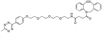
Methyltetrazine-PEG5-DBCO is a TCO reactive reagent with a DBCO group and water-soluble PEG spacer. This reagent can be used to convert azido-containing peptides or proteins into tetrazine-modified peptides or protein without catalyst or axillary reagents. DBCO is commonly used for copper-free Click Chemistry reactions. Reagent grade, for research purpose. Please contact us for GMP-grade inquiries.
Detail
Methyltetrazine-PEG5-DBCO is a TCO reactive reagent with a DBCO group and water-soluble PEG spacer. This reagent can be used to convert azido-containing peptides or proteins into tetrazine-modified peptides or protein without catalyst or axillary reagents. DBCO is commonly used for copper-free Click Chemistry reactions. Reagent grade, for research purpose. Please contact us for GMP-grade inquiries.
Other Products
Fungi/Yeast Genomic DNA Isolation Kits
Product Info
Document
Product Info
Overview
Rapid spin column purification of genomic DNA from viable yeast cells, fungal spores or mycelium, and bacteria including Gram-positive
Bead tubes (provided) allow for effective mechanical homogenization
Purified DNA is of high quality and integrity and compatible with any sensitive downstream applications such as PCR, qPCR, RFLP and more
These kits provide fast and reliable procedures for the purification of genomic DNA from viable yeast cells, fungal spores or mycelium, and bacteria including Gram-positive. Genomic DNA is efficiently extracted from the cells by a combination of heat treatment, detergents and Bead Tubes (provided). An optional lyticase treatment allows for improved DNA yields with certain fungal and yeast species. Recovered genomic DNA is of excellent yield and purity for any downstream application including PCR, qPCR, Restriction Fragment Length Polymorphism (RFLP), Amplified Fragment Length Polymorphism (AFLP), sequencing, SNP analysis and more.
Fungi/Yeast Genomic DNA Isolation Kit (Spin Column)
This kit provides rapid spin column purification of genomic DNA from viable yeast cells, fungal spores or mycelium, and bacteria including Gram-positive. Preparation time for a single sample is less then 30 minutes, and each kit contains sufficient materials for 50 preparations.
Fungi/Yeast Genomic DNA Isolation 96-Well Kit (HT)
Norgen’s Fungi/Yeast Genomic DNA Isolation 96-Well Kit provides a fast, reliable and simple procedure for high throughput isolation of DNA from viable yeast cells, fungal spores or mycelium and Gram-positive bacteria. The purification could be performed on either a vacuum manifold or using centrifugation. Complete 96 purifications in 40 minutes.
Maximum Amount of Starting Material: Fungi (Wet weight) Yeast or Gram-positive bacterial culture
50 mg 0.5 mL – 1 mL
Time to Complete 96 Purifications
40 minutes
Storage Conditions and Product Stability All solutions should be kept tightly sealed and stored at room temperature. These reagents should remain stable for at least 2 years in their unopened containers.
N-(t-Boc-Aminooxy-PEG2)-N-bis(PEG3-propargyl) is a branched crosslinker molecule with two terminal propargyl groups and a t-Boc protected aminooxy group. The propargyl groups can react with azide-bearing compounds or biomolecules via copper catalyzed azide-alkyne Click Chemistry to yield a stable triazole linkage. The protected amine can be deprotected under acidic conditions. Reagent grade, for research purpose. Please contact us for GMP-grade inquiries.
Document
N-(t-Boc-Aminooxy-PEG2)-N-bis(PEG3-propargyl) is a branched crosslinker molecule with two terminal propargyl groups and a t-Boc protected aminooxy group. The propargyl groups can react with azide-bearing compounds or biomolecules via copper catalyzed azide-alkyne Click Chemistry to yield a stable triazole linkage. The protected amine can be deprotected under acidic conditions. Reagent grade, for research purpose. Please contact us for GMP-grade inquiries.
The Opentrons Thermocycler Module GEN2 is a fully automated on-deck thermocycler, providing hands-free PCR in a 96-well plate format. Compatible with Opentrons hardware and software to allow for fully automated PCR reaction setup and thermocycling on your Opentrons robot. The heated lid and disposable seal fit tightly over the plate, ensuring efficient sample heating and minimal evaporation.
If you choose to include the Opentrons Flex Caddy and Calibration Adapter, your order will ship in 20 business days.
Document
The Opentrons Thermocycler Module GEN2 is a fully automated on-deck thermocycler, providing hands-free PCR in a 96-well plate format. Compatible with Opentrons hardware and software to allow for fully automated PCR reaction setup and thermocycling on your Opentrons robot. The heated lid and disposable seal fit tightly over the plate, ensuring efficient sample heating and minimal evaporation.
If you choose to include the Opentrons Flex Caddy and Calibration Adapter, your order will ship in 20 business days.