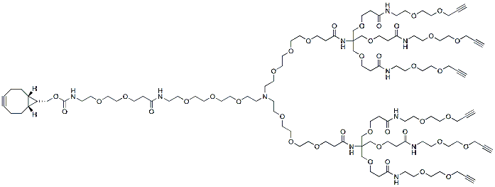
N-(endo-BCN-PEG2-amido-PEG3)-N-bis-(PEG3-Amino-Tri-(Propargyl-PEG2-ethoxymethyl)-methane) is is a multi-functional PEG linker with six terminal propargyl groups and a BCN group. The propargyl groups enables formation of triazole linkage with azide-bearing compounds or biomolecules in copper catalyzed Click Chemistry. The BCN group can react with azide -tagged compound or biomolecules. The hydrophilic PEG spacer increases solubility in aqueous media. Reagent grade, for research purpose. Please contact us for GMP-grade inquiries.
Detail
N-(endo-BCN-PEG2-amido-PEG3)-N-bis-(PEG3-Amino-Tri-(Propargyl-PEG2-ethoxymethyl)-methane) is is a multi-functional PEG linker with six terminal propargyl groups and a BCN group. The propargyl groups enables formation of triazole linkage with azide-bearing compounds or biomolecules in copper catalyzed Click Chemistry. The BCN group can react with azide -tagged compound or biomolecules. The hydrophilic PEG spacer increases solubility in aqueous media. Reagent grade, for research purpose. Please contact us for GMP-grade inquiries.
Other Products
DBCO-PEG4-triethoxysilane
Product Info
Document
Product Info
DBCO-PEG4-triethoxysilane is a PEG linker containing a triethoxysilane moiety and a DBCO group. Triethoxysilane is commonly used for surface modifications. DBCO group can react with azide-bearing compounds or biomolecules to form a stable triazole linkage without copper catalyst. The hydrophilic PEG chain increasse the water solubility of a compound in aqueous media. Reagent grade, for research purpose. Please contact us for GMP-grade inquiries.
Document
DBCO-PEG4-triethoxysilane is a PEG linker containing a triethoxysilane moiety and a DBCO group. Triethoxysilane is commonly used for surface modifications. DBCO group can react with azide-bearing compounds or biomolecules to form a stable triazole linkage without copper catalyst. The hydrophilic PEG chain increasse the water solubility of a compound in aqueous media. Reagent grade, for research purpose. Please contact us for GMP-grade inquiries.
The 50 bp DNA Ladder is prepared to ensure quality and batch-to-batch consistency. This Ladder contains ten discrete fragments ranging from 50 bp to 500 bp with a higher intensity reference band at 250 bp. This Ladder is ideal for quick sizing of PCR products
Contents 1mL of premixed DNA ladder (0.5µg/10µL) in loading buffer (10mM EDTA, 10% glycerol, 0.015% bromophenol blue, and 0.17% SDS).
Ladder Properties: • Ten discrete bands, ranging from 50 bp to 500 bp • Higher intensity band at 250 bp for easy reference
Fragment
Size (bp)
Mass (ng)
1
500
79
2
450
67
3
400
59
4
350
55
5
300
49
6
250
76
7
200
33
8
150
22
9
100
31
10
50
29
Recommended Use:
Mix thoroughly. For best results, load 10µL of DNA ladder per well. For precise mass determination with a densitometer, stain gel after electrophoresis using 0.5µg/mL ethidium bromide for 30-40 minutes. The table above shows the size and mass for each band based on 10µL ladder per well.
Storage:
Stable at room temperature. For longer term storage, -20°C is recommended.
This ladder was standardized using 10µL of DNA per lane on a 0.8 cm thick, 13 x 15 cm, 1.0% agarose gel run in TAE buffer.