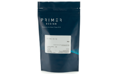
Primer and probe mix (150 reactions) Reverse Transcription, target specific primers (RNA genome viruses only) Copy number standard curve (sufficient for multiple standard curves) Internal extraction control – Read through VIC channel* Endogenous control (150 tests) RNAse/DNAse free water *alternative fluorophores available on request
Nucleic acid testing (NAT) is the method of choice for detection and quantification of a wide range of micro organisms. Primerdesign manufactures and supplies high quality quantitative real-time PCR kits for the detection and simultaneous quantification of numerous significant pathogens . A copy number standard curve is provided for quantification and an the internal extraction template (DNA or RNA), controls for the quality of the nucleic acid extraction and eliminates false negative results.
The kit is designed with the broadest possible detection profile to ensure that all clinically relevant strains and subtypes are detected. Target sequences are selected by working with data from key opinion leaders in the field. Multiple sequence alignments and unprecedented real-time PCR expertise in design and validation ensure the best possible kit. Details of the target and priming specificity are included in the individual handbooks above.
Packaged, optimised and ready to use. Expect Better Data.
Other Products
2 Layer Cell Factory One Wide Mouth And One Narrow Mouth
Product Info
Document
Product Info
2 Layer Cell Factory One Wide Mouth And One Narrow Mouth
Huayi Cell Factory is a robust and easy-to-use cell culture platform for applications in the production of human and animal vaccines, therapeutic proteins, cell therapy, and gene therapy.
To address the needs of your workflow, We have three kinds of mouth, which is available on most cell culture products to ensure consistent performance from lot to lot and from format to format.
The product assembled with ultrasonic welded technology
Versatile port design facilitates both pouring and aseptic filling techniques
Gamma radiation sterilization
the cell culture surface area
of one 10-layer Cell Factory unit is equivalent to the area
of 36 T-175 flasks
Document
Huayi Cell Factory is a robust and easy-to-use cell culture platform for applications in the production of human and animal vaccines, therapeutic proteins, cell therapy, and gene therapy.
To address the needs of your workflow, We have three kinds of mouth, which is available on most cell culture products to ensure consistent performance from lot to lot and from format to format.
Collagen is a fundamental component of the extracellular matrix, and the predominant protein in animals, constituting around 30% of total protein mass. A glycoprotein, it is well known for its triple helical structure. This is formed from three polypeptide α-chains with Gly-X-Y repeating residues (Gly for Glycine, X for proline, and Y for hydroxyproline).
Types of Collagen
Over 28 types of collagens have been identified, with Type I collagen being the most abundant. It’s prevalent in ligaments, tendons, skin, and bone tissue. Its mature, insoluble form grants it remarkable strength, making it vital for the mobility of organisms. Collagen also has biochemical functions, influencing cell growth, proliferation, and differentiation.
This version of the kit is designed to detect and measure INSOLUBLE forms of collagen. Chose our Sircol 2.0 collagen kit if you need to analyse SOLUBLE collagen.
Applications of Collagen
Collagen, with its diverse properties, finds utility in various industries. It plays a role in medicine for wound healing and has an expanding role in tissue engineering and cell culture for biomedical purposes. It’s gaining popularity in the cosmetic industry for skin rejuvenation and is used in chemical formulations and the food industry as a functional food supplement and additive.
How does the Sircol assay detect collagen?
Sircol dye reagent contains Sirius Red – a linear anionic dye with sulphonic acid side chain groups. Under assay conditions the Sircol dye binds the basic groups of soluble collagen molecules. Maximal binding occurs in collagens possessing intact triple helix organisation as the highly ordered Gly-X-Yn helical structure of tropocollagen further contributes to dye binding. This results in a high degree of dye-collagen specificity. Affinity is progressively reduced during heat denaturation 4ºC due to the unwinding of the triple helix and formation of random chains.
Overview of the Sircol assay process:
Step 1. Samples being assayed for insoluble collagen must first undergo a 2-3 hour pre-treatment with Sircol Fragmentation reagent. This converts insoluble collagen into water-soluble gelatin can then be assayed.
Step 2. Addition of Sircol Dye Reagent to these pre-treated insoluble collagen samples results in the formation of a denatured collagen-dye complex. This complex then precipitates during the dye incubation period and is subsequently isolated by centrifugation, followed by washing to remove unbound dye. The Denatured collagen-bound dye is then eluted and measured spectrophotometrically.
Step 3. The insoluble collagen content of unknown samples is quantified by comparison against a calibration curve prepared using a the denatured collagen standard supplied with the kit.
Assay range
100 – 1000 µg/ml
Limit of Detection
100µg/ml
Detection Method
Colorimetric Detection (556nm) (Endpoint)
Measurements per kit
110 in total (allows a maximum of 46 samples to be run in duplicate alongside a standard curve).
Suitable Samples
The assay can be used to assess the rate of production of newly laid down collagen fibres during periods of rapid growth, development, tissue repair, remodeling and wound healing. Sources of material includes tissues, bone and calcified tissue.
*Insoluble collagens must be converted into soluble form prior to assay. Instructions and regents are provided with the kit., depending on sample this will require prior salt/acid/acid-pepsin extraction.
**non-mammalian collagens may result in a reduced limit of detection. We recommend use of an assay standard matched to the species under assay.
Many customers have found that the straightforward sample processing and analysis of Sircol make it a good alternative to conventional hydroxyproline analysis.
Precautions
This kit is designed for research use only. Not for use in diagnostic procedures. Kit requires access to a centrifuge, water bath / heated block, as well as a spectrophotometer/colorimeter capable of absorbance detection at 556nm. Specific sample preparation protocols may require customer to provide further reagents, consult assay manual for further information.
Sircol Insoluble Collagen kit contents:
1. Sircol Dye Reagent (1x110ml)
2. Denatured Collagen Reference Standard (1x5ml, 1.0mg/ml)
3. Acid-Salt Wash Reagent (1x20ml)
4. Fragmentation Reagent (1x110ml)
5. Alkali Reagent (1x110ml)
6. 2ml screw-cap tubes for preparation of samples.
7. Assay kit manual
NB: Additional reagents may be required for sample preparation prior to assay. Consult manual or contact us for further details.
Document
As collagens mature, they become increasingly crosslinked and insoluble – characteristics necessary for key biophysical role that collagen plays in living organisms. Biocolor’s Sircol™ INSOLUBLE Collagen Kit is a dye-binding assay designed for accurate quantification and measurement such collagens. It is ideal for analyzing crosslinked / insoluble collagens from sources such as tissues, bone, and calcified tissue.
Attogene has developed the first of its kind and patent pending technology that uses a novel RNA/DNA substrate tagged on the 5′ and 3′ end with a Biotin that is attached to our streptavidin colloidal gold reporter molecules and a test line tag. In the absence of RNases H, the gold tagged RNA/DNA molecule will flow up the lateral flow strip and bind to the test line using an anti tag antibody (INDICATING that NO RNase H is detected). When RNases H is present or added, however, the RNA strand of the RNA/DNA substrate is cleaved, and the Biotin tagged 5′ and 3′ tags are spatially separated in solution cauing the test line to disapear.
This test can be used to rapidly and efficiently detect ribonucleases H activity and is a perfect tool for monitoring unit activity or residual RNAse H activity.
Document
RNase H activity in a convenient and sensitive colormetric assay that delivers results in real time. Great for Quality Testing for RNase H activity in enzyme preparations and determination of RNase H unit activity.