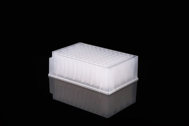
NEST 2 mL 96-Well Deep Well Plate, V Bottom (50 count)
Facebook
X
Pinterest
Email
NEST 2 mL 96-Well Deep Well Plate, v-bottom, polypropylene, sterile, DNase, RNase, pyrogen and endotoxin free
Conforms to SBS and ANSI requirements
Centrifugal strength up to 3000g
Square well
50 count
Labware definition is available for immediate use in Opentron’s Labware Library
Detail
NEST 2 mL 96-Well Deep Well Plate, v-bottom, polypropylene, sterile, DNase, RNase, pyrogen and endotoxin free
Conforms to SBS and ANSI requirements
Centrifugal strength up to 3000g
Square well
50 count
Labware definition is available for immediate use in Opentron’s Labware Library
Other Products
CloneSizer 100 bp DNA Ladder
Product Info
Document
Product Info
Overview
Ready-to-use
Quantitative
Highly Stable
Precise
Fourteen discrete fragments ranging from 100 bp to 2000 bp in 100 bp increments
Higher intensity reference bands at 500 bp and 1000 bp
2686 bp (pUC19) reference band for easy clone identification
The CloneSizer 100 bp DNA Ladder is prepared to ensure quality and batch-to-batch consistency. Our CloneSizer contains fourteen discrete fragments ranging from 100 bp to 2000 bp in 100 bp increments, double intensity reference bands at 500 and 1000 bp and an additional 2686 bp (pUC19) reference band for easy clone identification.
Contents: 1mL of premixed DNA ladder (0.5µg/10µL) in loading buffer (10mM EDTA, 10% glycerol, 0.015% bromophenol blue, and 0.17% SDS).
CloneSizer 100 bp DNA Ladder (Cat# 11600) – 100 loads
Ladder Properties:
Fourteen discrete fragments ranging from 100 bp to 2000 bp in 100 bp increments
Higher intensity reference bands at 500 bp and 1000 bp
2686 bp (pUC19) reference band for easy clone identification
Fragment
Size (bp)
Mass (ng)
1
2686
72
2
2000
53
3
1500
41
4
1200
42
5
1000
56
6
900
30
7
800
29
8
700
25
9
600
25
10
500
52
11
400
19
12
300
20
13
200
19
14
100
17
Recommended Use:
Mix thoroughly. For best results, load 10µL of DNA ladder per well. For precise mass determination with a densitometer, stain gel after electrophoresis using 0.5µg/mL ethidium bromide for 30-40 minutes. The table above shows the size and mass for each band based on 10µL ladder per well.
Storage:
Stable at room temperature. For longer term storage, -20°C is recommended.
This ladder was standardized using 10µL of DNA per lane on a 0.8 cm thick, 13 x 15 cm, 1.0% agarose gel run in TAE buffer.
Widely used in the area of laboratory, pharmaceutical factory, biochemistry, biological products and so on. It is the ideal instruments for blood separation, protein precipitation and cell collection.
1. Brushless DC motorin great torque, free maintenance, no carbon dust pollution, quick in speed up and down. 2. Flexible axledriven system which drive the rotor directly, smooth in operation, low noise and small vibration. 3. Automatically electric lid lock, super speed, over temperature protection and imbalance protection. 4. Microprocessor control, speed, time, temperature and RCF in operation, speed rising and reducing quick, operate simply. 5.10 kinds of program storedin the memory, 10 kinds of accelerating and decelerating speed for your choice. 6. There are many rotors for your choice, adapters are available by experiment requirements. 7.3 tiers protection steel cover, safe and reliable.
Technical Parameters
for 6000rpm 4x1000ml 6x500ml 6x300ml Angle Rotor Laboratory Centrifuge Machine
Max Speed: 6000r/min
Max.RCF: 6600xg
Max Volume: 4x1000ml
Power supply:AC220V,50HZ,10A
Timer: 1~99min
Noise:≤55dBA
Dimension:800×645×830mm
Net Weight: 170KG
Speed Accuracy: ±20r/min
Package(W*D*H): 920×800×1320mm
Temperature range:
Inapplicable
Matched Rotorsfor DD6 laboratory centrifuge
Order No.
Rotor type
Max.Speed (rpm)
Max.Volume (ml)
Max.RCF (xg)
No33097
Swing out rotor
4,000
4x1000ml (round/square cup )
4,060
No30221
Angle rotor
6,000
4x300ml
5,390
No30222
Angle rotor
6,000
6x300ml
5,660
No30223
Angle rotor
6,000
6x500ml
6,600
Packing & Shipping_____________________
by Sea/ by Air / by Express Sevices DHL, Fedex, TNT…
Document
DD6 is floor standing large capacity 4x1000ml, 6x300ml, 6x500ml laboratory centrifuge machine without refrigerated one. It is up to max.speed 6000rpm.
Q-PAGE™ Bis-Tris Precast Gel is a high-performance and easy to use precast polyacrylamide gel for electrophoresis in Bis-Tris buffer system (MOPS or MES). The optimized gel formula allows Q-PAGE™ Bis-Tris Precast Gel to show improved resolution, accurate results, and an extended shelf-life over conventional Laemmli Tris-HCl gels.
Q-PAGE™ Bis-Tris Precast Gels are available in gradient (4 to 12%) and fixed (8% and 12%) concentrations of polyacrylamide in 12-and 15-well formats. Two available cassette sizes, Mini (10 x 8.3 cm) and Midi (10 x 10 cm), are compatible with most popular protein electrophoresis systems. Q-PAGE™ Mini (QP2XXX) Gels are suitable for Bio-Rad® and other systems. Q-PAGE™ Midi (QP3XXX) Gels are suitable for Invitrogen® XCell SureLock® Mini-Cell, Invitrogen® Mini Gel Tank, Hoefer SE260, and other systems.
Key Features
User-friendly gel cassette:
Numbered and framed wells for sample loading
Labeled warning sign and green tape as reminder
Enhanced gel performance:
Enhanced band sharpness
Better resolution of small proteins
Stable for shipping at ambient temperature
Easy compatibility:
Available as homogeneous and adjusted gradient gels for a wide range of protein separation.
Compatible with most popular protein electrophoresis systems
Storage and stability
Store Q-PAGE™ Precast Gels at 4°C for periods up to 12 months.
Do not freeze Q-PAGE™ Precast Gels. Remove tape and comb before electrophoresis.
Keep Q-PAGE™ Precast Gels flat during storage.
Document
Q-PAGE™ Bis-Tris Precast Gel is a high-performance and easy to use precast polyacrylamide gel for electrophoresis in Bis-Tris buffer system (MOPS or MES). The optimized gel formula allows Q-PAGE™ Bis-Tris Precast Gel to show improved resolution, accurate results, and an extended shelf-life over conventional Laemmli Tris-HCl gels.
Q-PAGE™ Bis-Tris Precast Gels are available in gradient (4 to 12%) and fixed (8% and 12%) concentrations of polyacrylamide in 12-and 15-well formats. Two available cassette sizes, Mini (10 x 8.3 cm) and Midi (10 x 10 cm), are compatible with most popular protein electrophoresis systems. Q-PAGE™ Mini (QP2XXX) Gels are suitable for Bio-Rad® and other systems. Q-PAGE™ Midi (QP3XXX) Gels are suitable for Invitrogen® XCell SureLock® Mini-Cell, Invitrogen® Mini Gel Tank, Hoefer SE260, and other systems.