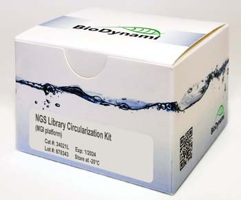
The NGS Library Circularization Kit (MGI Platform) was developed for preparation of single-stranded circular DNA libraries for next generation sequencing (MGI platform).
Detail
The NGS Library Circularization Kit (MGI Platform) was developed for preparation of single-stranded circular DNA libraries for next generation sequencing (MGI platform).
The kit uses linear dsDNA libraries (MGI platform) as input and enriches the circularized single-stranded DNA libraries. The circularization kit has a higher library circularization efficiency (25%) as compared to other vendors (7-15%).
NGS Library Circularization Kit workflow
Comparison of Library Circularization Efficiency
Kit features
Fast protocol
Total protocol time is around 1 hour
Hands-on time is only ~10 minutes
Guaranteed high library circularization efficiency as compared to other kits
t-Boc-N-amido-PEG4-Amide-Tri-(propargyl-PEG10-ethoxymethyl)-methane is a branched crosslinker molecule with three terminal propargyl groups and a t-Boc protected amide group. The propargyl groups can react with azide-bearing compounds or biomolecules via copper catalyzed azide-alkyne Click Chemistry to yield a stable triazole linkage. The protected amine can be deprotected under acidic conditions. Reagent grade, for research use only.
Document
t-Boc-N-amido-PEG4-Amide-Tri-(propargyl-PEG10-ethoxymethyl)-methane is a branched crosslinker molecule with three terminal propargyl groups and a t-Boc protected amide group. The propargyl groups can react with azide-bearing compounds or biomolecules via copper catalyzed azide-alkyne Click Chemistry to yield a stable triazole linkage. The protected amine can be deprotected under acidic conditions. Reagent grade, for research use only.
Gel images of different ranges of library size selection. Sheared human genomic DNA was used as input.
.
Library size selection is an enrichment of a specific range of library sizes for NGS library preparations. The NGS library preparation is related to the quality of the sequencing data. Precise NGS library size selection can increase sequencing efficiency, improve data quality, and reduce costs.
There are two types of sequencing technologies: short-read sequencing and long-read sequencing. Short-read sequencing uses DNA libraries that contain small insert DNA fragments of similar sizes, usually several hundred base pairs. The sequencing efficiency can be improved if the DNA size selection is in the right range. Cat.# 20104S and 20104L are the best kits for NGS library size selection of illumina paired-end 100 (PE100) sequencing with 100-200 bp library inserts; Cat.# 20105S and 20105L are the best kits for NGS library size selection of illumina paired-end 150 (PE150) sequencing with 150-300 bp library inserts; and Cat.# 20106S and 20106L are the best kits for NGS library size selection of illumina paired-end 300 (PE300) sequencing with 300-600 bp library inserts.
Long-read sequencing uses a large DNA fragment as input and makes very long reads. Usually, library size selection is preferred to remove smaller fragments. Cat.# 20110S and 20110L are the best kits for long-read sequencing size selection with DNA sizes >5 kb, and Cat.# 20111S and 20111L are the best kits for long-read sequencing size selection with DNA sizes >10 kb.
The magnetic beads, or SPRI (Solid Phase Reversible Immobilization) beads, is well used for the purification of DNA due to their reversible DNA binding. The NGS library can be size-selected by the magnetic beads or SPRI beads. The properties of the magnetic beads can be changed for a specific range of DNA binding. The contaminants and other unwanted components in the libraries can also be removed during size selection.
Specific ranges of NGS libraries can be selected using magnetic beads with different buffer compositions. The first DNA-beads binding step, also called the right-side clean-up, removes large NGS library fragments. The large NGS library fragments that bind to the beads are discarded with the beads pellet. The desired NGS library fragments in the supernatant are transferred to a new well, and new beads are added to the supernatant for the second beads-DNA binding, also called the left-side clean-up. After the rinsing step, the NGS library fragments with the dual selection are eluted in water or an appropriate buffer. The magnetic beads method has great advantages over time-consuming column purification and tedious gel-based purification.
NGS library size selection with dual clean-ups.
.
Library size selection for long-read sequencing only requires a single clean-up. In this case, only the large library fragments are bound to the beads, while other small library fragments are discarded with the supernatant. The selected larger library fragments are eluted in water or an appropriate buffer after the rinsing step.
NGS library size selection with single clean-up for >5 kb and >10 kb libraries.
[CD1021] SMOChem™ Deoxynucleotide (dNTP) Mix, 25 mM each (100 mM total), 500 µl x 6
Product Info
Document
Product Info
Description
The SMOChem™ Deoxynucleotide (dNTP) Mix is an aqueous solution that contains an equimolar solution of ultrapure dATP, dCTP, dGTP and dTTP, each at a concentration of 25 mM at pH 8.5. The dNTP Mix is designed for many different molecular biology applications that involved in DNA synthesis or labeling, such as PCR, real-time PCR, DNA sequencing, reverse transcription, primer extension, and etc. The dNTP Mix is free of exo-deoxyribonuclease and endo-deoxyribonuclease as well as ribonuclease activity. The dNTP Mix offers the possibility to reduce the number of pipetting steps and the risk of reaction set up errors.
Features
Ideal for PCR amplification and cDNA synthesis
Premixed solution
Nuclease and ribonuclease free
Applications
PCR amplification of DNA fragments
DNA fill-in reaction
DNA sequencing
Reverse transcription
One-step RT-PCR
Storage
-20°C for 36 months
Document
The SMOChem™ Deoxynucleotide (dNTP) Mix is an aqueous solution that contains an equimolar solution of ultrapure dATP, dCTP, dGTP and dTTP, each at a concentration of 25 mM at pH 8.5. The dNTP Mix is designed for many different molecular biology applications that involved in DNA synthesis or labeling, such as PCR, real-time PCR, DNA sequencing, reverse transcription, primer extension, and etc. The dNTP Mix is free of exo-deoxyribonuclease and endo-deoxyribonuclease as well as ribonuclease activity. The dNTP Mix offers the possibility to reduce the number of pipetting steps and the risk of reaction set up errors.