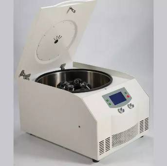
Oil test centrifuge, Heating oil test centrifuge, special tube oil test centrifuge
Detail
Features:
1. Programmable microprocessor control system, touch screen indicates rotor No., speed, time, temperature, RCF, rate of speed as well as fault information.
2. Adopts band heater, fast heating speed, temperature uniform, with the function of temperature-control and constant.
3. Centrifugal tube made in special skill, anti-broken when work.
4. Great toque brushless motor, maintenance-free, no powder, fast acceleration and deceleration, low noise, which improve the stability a lot.
5. Electric lid lock, over-speed and over-temperature protection
6.3 phase to keep the best performance.
7. Stainless steel chamber, safe and reliable
TD5B Technical Parameter:
Max. Speed
4000rpm
Max. RCF
2498×g
Max. Capacity
4×100ml
Time Range
0~99min
Rotor
Swing Out Rotor
Noise (dB)
≤ 58
Temperature
Room temperature:+10~80℃
Acc/Dcc
10 Kinds
Speed Accuracy
±20r/min
Temperature Accuracy
±1℃
Oil tube size
37×200mm
Size (W x D x Hmm)
685×500×385mm
Net Weight(Kg)
67KG
Other Products
Heat Labile Exonuclease I
Product Info
Document
Product Info
OverView
Heat-Labile Exonuclease I (HL-ExoI) is a 3’ – 5’ exonuclease, specific for single stranded DNA. The enzyme is recombinantly produced in E. coli. HL-ExoI is active at 25 – 37°C and inactivated by 1 minute incubation at 80°C or 15 minutes at 60°C.
HL-ExoI is used for degradation of ssDNA such as primers and oligos. It is also ideal for treatment of sensitive samples and useful in the development of novel molecular diagnostics applications.
Key Features
3’-5’ exonuclease specific for single stranded DNA
High activity at 25 – 37°C
Easily heat-inactivated by 1 min incubation at 80°C, or 15 min at 60°C
Moderate salt tolerance
Application
Removal of primers post-PCR prior to DNA sequencing or SNP detection
Figures
Properties
Quality Control
ArcticZymes is dedicated to the quality of our products. We manufacture all products at our ISO 13485 certified facility in Norway.
Document
Heat-Labile Exonuclease I (HL-ExoI) is a 3’ – 5’ exonuclease, specific for single stranded DNA. The enzyme is recombinantly produced in E. coli. HL-ExoI is active at 25 – 37°C and inactivated by 1 minute incubation at 80°C or 15 minutes at 60°C.
Q-PAGE™ TGN (Tris-Glycine Novel) Precast Gels are ready-to-use acrylamide gels for SDS-PAGE running in Tris-Glycine buffer system. With unique formula, Q-PAGE™ TGN Precast Gels perform enhanced speed, better separation, and longer shelf life as compared with conventional Laemmli Tris-HCl gels. The protein migration patterns in Q-PAGE™ TGN series, however, are similar with typical Laemmli Tris-HCl gels, and thus Q-PAGE™ TGN Precast Gels are compatible to traditional SDS-PAGE and subsequent analyses.
Q-PAGE™ TGN Precast Gels are available in gradient (4 to 15%) and fixed (10%) concentrations of polyacrylamide in 12- and 15-well formats. Two available cassette sizes, Mini (10 x 8.3 cm) and Midi (10 x 10 cm), are compatible with most popular protein electrophoresis systems. Q-PAGE™ Mini (QP4XXX) Gels are suitable for Bio-Rad® and other systems. Q-PAGE™ Midi (QP5XXX) Gels are suitable for Invitrogen® XCell SureLock® Mini-Cell, Invitrogen® Mini Gel Tank, Hoefer SE260, and other systems.
Key Features
User-friendly gel cassette:
Numbered and framed wells for sample loading
Labeled warning sign and green tape as reminder
Enhanced gel performance:
Enhanced gel electrophoresis speed
Better band separation
Stable for shipping at ambient temperature
Easy compatibility:
Available as homogeneous and adjusted gradient gels for a wide range of protein separation.
Compatible with most popular protein electrophoresis systems
Storage and stability
Store Q-PAGE™ Precast Gels at 4°C for periods up to 12 months.
Do not freeze Q-PAGE™ Precast Gels. Remove tape and comb before electrophoresis.
Keep Q-PAGE™ Precast Gels flat during storage.
Document
Q-PAGE™ TGN (Tris-Glycine Novel) Precast Gels are ready-to-use acrylamide gels for SDS-PAGE running in Tris-Glycine buffer system. With unique formula, Q-PAGE™ TGN Precast Gels perform enhanced speed, better separation, and longer shelf life as compared with conventional Laemmli Tris-HCl gels. The protein migration patterns in Q-PAGE™ TGN series, however, are similar with typical Laemmli Tris-HCl gels, and thus Q-PAGE™ TGN Precast Gels are compatible to traditional SDS-PAGE and subsequent analyses.
Q-PAGE™ TGN Precast Gels are available in gradient (4 to 15%) and fixed (10%) concentrations of polyacrylamide in 12- and 15-well formats. Two available cassette sizes, Mini (10 x 8.3 cm) and Midi (10 x 10 cm), are compatible with most popular protein electrophoresis systems. Q-PAGE™ Mini (QP4XXX) Gels are suitable for Bio-Rad® and other systems. Q-PAGE™ Midi (QP5XXX) Gels are suitable for Invitrogen® XCell SureLock® Mini-Cell, Invitrogen® Mini Gel Tank, Hoefer SE260, and other systems.
endo-BCN-PEG3-Gly represents an ADC linker featuring a BCN group, a hydrophilic PEG3 linker, and glycine residue. The BCN group is a click chemistry handle that readily reacts with azide groups on a target molecule to form stable triazole linkages. The PEG units enhance the aqueous solubility of the compound and may assist in fine-tuning DMPK properties. This compound may be applied to the synthesis of antibody-drug conjugates.
Document
endo-BCN-PEG3-Gly represents an ADC linker featuring a BCN group, a hydrophilic PEG3 linker, and glycine residue. The BCN group is a click chemistry handle that readily reacts with azide groups on a target molecule to form stable triazole linkages. The PEG units enhance the aqueous solubility of the compound and may assist in fine-tuning DMPK properties. This compound may be applied to the synthesis of antibody-drug conjugates.