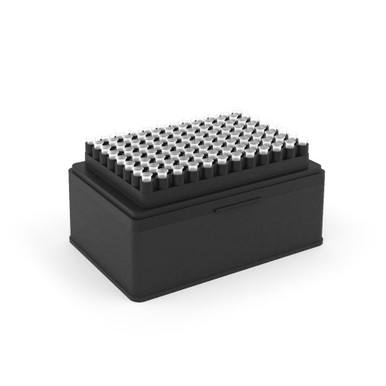
Opentrons OT-2 Tips, 1000µL. Optimized for volumes 100µL to 1000µL Clear Tips, Polypropylene. These tips are DNAse/RNAse, pyrogen and protease free – they are sterilized from ebeam irradiation. Not autoclavable.
Opentrons OT-2 Tips, 1000µL. Optimized for volumes 100µL to 1000µL Clear Tips, Polypropylene. These tips are DNAse/RNAse, pyrogen and protease free – they are sterilized from ebeam irradiation. Not autoclavable.
For selective enrichment culture of Listeria monocytogenes in food.
Principle and Interpretation
Tryptone, proteose peptone, meat extract, yeast paste powder provide nitrogen sources, vitamins, amino acids, and growth factors; sodium chloride can maintain a balanced osmotic pressure; sodium dihydrogen phosphate and potassium dihydrogen phosphate as buffering agents; esculin are fermentable sugars; lithium chloride and other antibiotics can inhibit Gram negative bacteria and most Gram positive bacteria.
Formulation
Ingredients
/liter
Enzymatic digest of animal tissues ( Proteose peptone)
5.0g
Enzymatic digest of casein ( Tryptone)
5.0g
Meat extract
5.0g
Yeast extract
5.0g
Sodium chloride
20.0g
Disodium hydrogen phosphate dihydrate
12.0g
Potassium dihydrogen phosphate
1.35g
Aesculin
1.0g
Lithium chloride
3.0g
pH7.2±0.2 at 25°C
Preparation
Suspend 57.4g in 1 L of distilled or deionized water. Heat with frequent agitation and boil to completely dissolve the powder. Distribute into flasks. Autoclave at 121℃ for 15 minutes. Cool to 45-50℃,aseptically add the contents of 1 vial of Fraser Selective Supplement (SR0120) to each 225 mL of base to prepare FB1 solution; Add one reagent (SR0130) to each100 mL of FB2 culture medium to prepare FB2 enrichment solution.
Quality Control
Cultural characteristics observed after incubation at 35-37°C for 46~50 hours
2-30℃,Keep container tightly closed, avoid direct sunlight.
Use before expiry date on the label.
Precautions
1. When weighing the dehydrated medium, please wear masks to avoid causing respiratory system discomfort
2. Keep container tightly closed after using to prevent clumping.
Waste Disposal
Microbiological contamination was disposed by autoclaving at 121°C for 30 minutes.
Revision
On June 14, 2024
References
ISO 11290
Document
Intended Use For selective enrichment culture of Listeria monocytogenes in food. Principle and Interpretation Tryptone, proteose peptone, meat extract, yeast paste powder provide nitrogen ……
DBCO-Acid is a non-activated building block and adds minimal spacer to modified molecules. In the presence of activators such as EDC or HATU, this reagent can be used to derivatize amine-containing molecules through a stable amide bond. DBCO is commonly used for copper-free Click Chemistry reactions. Reagent grade, for research purpose. Please contact us for GMP-grade inquiries.
Document
DBCO-Acid is a non-activated building block and adds minimal spacer to modified molecules. In the presence of activators such as EDC or HATU, this reagent can be used to derivatize amine-containing molecules through a stable amide bond. DBCO is commonly used for copper-free Click Chemistry reactions. Reagent grade, for research purpose. Please contact us for GMP-grade inquiries.
Champion™ Competent Cells are chemically competent cells, which were prepared by SMOBIO to make E. coli perform excellent transformation efficiency. Standard transformation protocol is recommended for large plasmids or non-ampicillin selection. Time-saving transformation protocol is recommended for simple and rapid transformation. Champion™ Competent Cells are one of the fastest and simplest ready-to-use competent cell products in the world.
Kit contents
Champion™ Competent Cells
pUC19 Control Plasmid (5 μl, 10-4 μg/μl)
Champion™ Transformation Protocol Card
Shipping condition
Throughout the shipping process, the temperature is maintained under -70°C.
Storage and expiration
Champion™ Competent Cells must be stored between -70°C to -80°C. Subsequent freeze-thaw cycles will reduce transformation efficiency. If high efficiency is required for the experiment, do not use aliquots that have gone through several freeze-thaw cycles. The efficiency of Champion™ Competent Cells lasts for 1 year with proper storage.
Document
Champion™ Competent Cells are chemically competent cells, which were prepared by SMOBIO to make E. coli perform excellent transformation efficiency. Standard transformation protocol is recommended for large plasmids or non-ampicillin selection. Time-saving transformation protocol is recommended for simple and rapid transformation. Champion™ Competent Cells are one of the fastest and simplest ready-to-use competent cell products in the world.