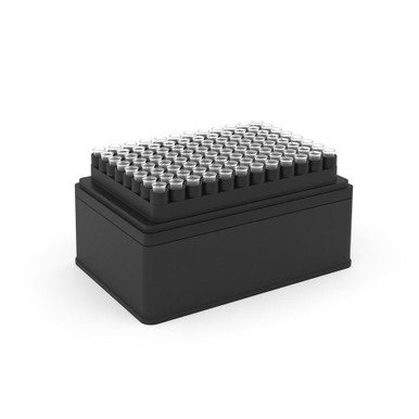
Opentrons OT-2 Tips, 300µL. Optimized for volumes 5µL to 300µL Clear Tips, Polypropylene. These tips are DNAse/RNAse, pyrogen and protease free – they are sterilized from ebeam irradiation. Not autoclavable.
Apoptosis is an essentially normal physiological process that removes now redundant, cells, particularly during embryonic development and early growth. In adult animals the process removes cells that are irreparable. The apoptotic process is also involved in many major diseases such as cancer, where transformed tumour cells have their apoptotic process disabled, permitting cell cycling to continue unchecked. In contrast some forms of senile dementia may result from excessive apoptotic induction of neural cells.
The apoptotic process in mammalian cells is a rapid event (2‐4 hours). Within this short time span an apparently viable cell can be quietly dismantled, to disappear leaving no visible trace of its former existence.
How is apoptosis detected or measured?
An apoptosis cascade of activators, effectors and regulators has been identified. This in turn led to a range of apoptosis assays being devised to detect and monitor these events. Some laboratories will employ two distinct assays, one selected to detect early (initiation) apoptotic events, while a second assay will target a later (execution) event. Apoptosis assays, based on methodology, can be classified into four major inter‐linked groups:
[1] DNA fragmentation (electrophoresis and nick end labelling, TUNEL).
[2] Apoptotic proteases (fluorescently labelled antibodies to the caspases).
[3] Flow cytometric analysis (FACS, incorporating other group assays).
Biocolor’s APOPercentage assay is based on the latter. Further information can be found under the ‘Mode of Action’ Tab.
How does APOPercentage detect apoptosis?
The mammalian cell membrane has been described as a semi‐fluid mosaic structure, composed of phospholipids with a diverse group of inserted proteins and some cholesterol. The phospholipids are the major components of the membrane and are arranged in the form of a ‘bi‐layer’; which is asymmetric in composition, structure, and function.
To ensure normal transmembrane functions the phospholipids must be maintained in an asymmetric composition. The process is regulated by ‘flippases’, which catalyse the active transport of aminophospholipids from the outer to inner monolayer. However, in cells undergoing apoptosis, flippase is overwhelmed by the action of another enzyme, termed ‘floppase’ or ‘scramblase’. The net effect is a scrambling of the phospholipid distribution between the inner and outer monolayers.
Cell membrane changes during apoptosis
The APOPercentage assay utilises an intense, pink-coloured dye reagent which is taken up during in-vitro culture by apoptosis-committed cells. This uptake occurs at the stage of Phosphatidylserine transmembrane movement, as produced by the flipflop mechanism. Dye uptake continues until blebbing occurs. No further dye can then enter the now defunct cell and the dye that has accumulated within the cell is not released (unlike necrotic cells which release dye).
Since the dye reagent is excluded or not retained by healthy or necrotic cells it therefore acts as a specific label for apoptotic cells.
How are APOPercentage-labelled cells quantified?
Labelled apoptosis cells may then by conveniently analysed by the following methods:
Direct Analysis The intense pink colour of the labelled cells can be visually assessed using brightfield microscopy. Apoptosis in substrate-adherent cell populations is therefore readily quantified using image analysis techniques. This technique is the most sensitive with the ability of detecting one single apoptotic cell per well.
Colorimetry protocol Dye that accumulates within apoptotic cells is released into solution via addition of Dye Release Reagent. The concentration of this intracellular dye is then measured at 550nm using a microplate colorimeter/spectrophotometer.
NB: The APOPercentage assay kit does NOT require the use of a Flow Cytometer.
Limit of Detection
A single cell (via image analysis method)
Detection Method
Colorimetric (550nm) (Endpoint) or Image Analysis based
Measurements per kit
Sufficient for 4×24 well plates or 6×96 well plates
Suitable Samples
Adherent mammalian cells (in-vitro)
APOPercentage kit contents:
1. APOPercentage Dye (1x5ml)
2. Dye Release Reagent (1x150ml)
3. Phosphate Buffered Saline (PBS) (1x120ml)
4. 24-well starter plate.
5. Assay kit manual.
The Colorimetric Protocol requires a Microplate Colorimeter / Spectrophotometer.
Additional 96-well plates will be required for use when reading dye absorbance values.
The Direct Detection Protocol Requires an inverted stage microscope with an attached digital camera.
NB: Additional reagents (typically culture medium and suitable apoptosis treatments) may be required for sample preparation prior to assay. Consult manual or contact us for further details.
Document
The APOPercentage™ Apoptosis kit is a dye-based, colorimetric assay for detection and measurement of apoptosis (programmed cell death) during in-vitro cell culture.
This product allows rapid and reliable isolation of high-quality genomic DNA from various soil, stool, and other environmental samples. Up to 500mg soil, 100mg stool, or 0.5g environmental samples can be processed in 60 minute. Purified DNA is suitable for PCR, restriction digestion, and next-generation sequencing.
Details
Specifications
Features
Specifications
Main Functions
Isolation total DNA from 250-500mg soil sample
Applications
PCR, southern blot and enzyme digestion, etc.
Purification technology
Magnetic beads technology
Process method
Manual or automatic
Sample type
Soil
Sample amount
250-500mg
Elution volume
≥50μl
Time per run
≤70 minutes
Principle
This product is based on the purification method of high binding magnetic particles. Soil sample is homogenized and then treated in a specially formulated buffer containing detergent to lyse bacteria, yeast, and fungal samples. Humic acid, proteins, polysaccharides, and other contaminants are removed using our propietary Absorber Solution. After adding magnetic particles and binding solution, DNA will be adsorbed on the surface of magnetic particles, and impurities such as proteins will be removed without adsorption. The adsorbed particles were washed with washing buffer to remove proteins and impurities, washed with ethanol to remove salts, and finally DNA was eluted by Elution Buffer.
Advantages
High concentration – maximum extraction of total DNA from soil samples
High purity – purified DNA can be directly used in various downstream applications
Kit Contents
Contents
D635601
D635602
D635603
Purification Times
48 Preps
96 Preps
5 x 96 Preps
MagPure Particles
2.5 ml
5 ml
22 ml
2ml Bead Tubes
48
96
400
Buffer SOL
60 ml
100 ml
500 ml
Buffer SDS
6 ml
10 ml
50 ml
Reagent DX
1 ml
1 ml
5 ml
Buffer PS
10 ml
20 ml
90 ml
Absorber Solution
10 ml
20 ml
90 ml
Buffer GDP
70 ml
150 ml
2 x 350 ml
Buffer GW1*
22 ml
44 ml
220 ml
Elution Buffer
20 ml
20 ml
60 ml
Storage and Stability
MagPure Particles and Absorber Solution should be stored at 2-8°C upon arrival. However, short-term storage (up to 12 weeks) at room temperature (15-25°C) does not affect their performance. The remaining kit components can be stored dry at room temperature (15-25°C) and are stable for at least 18 months under these conditions.
Experiment Data
Document
This product allows rapid and reliable isolation of high-quality genomic DNA from various soil, stool, and other environmental samples. Up to 500mg soil, 100mg stool, or 0.5g environmental samples can be processed in 60 minute. Purified DNA is suitable for PCR, restriction digestion, and next-generation sequencing.