Plasmid MiniPrep Kit & High Throughput Kit (Magnetic Beads)
Facebook
X
Pinterest
Email
Detail
Plasmid MiniPrep Kit & High Throughput Kit (Magnetic Beads)
The kits were developed for plasmid miniprep directly from bacterial cultures. 100% centrifuge-free; NO vacuum needed; No column needed.
Plasmid MiniPrep Kit (Magnetic Beads), Cat.# 50012 – Use 2 ml of bacteria culture directly
Plasmid MiniPrep High Throughput Kit (Magnetic Beads), Cat.# 50011 – High throughput: Use 0.2 ml of bacteria culture directly with 96-well plates – Low throughput: Use 0.2 ml of bacteria culture directly with 1.5 ml tubes
The kits can be used for both high copy and low copy numbers of plasmid DNA. With BioDynami’s proprietary magnetic beads technology, the kits eliminate the requirement of centrifuge, vacuum, and column. Our magnetic beads provide a robust and reliable tool for both high throughput and low throughput applications of plasmid DNA isolation with high binding capacity and fast magnetic response time.
The High Through kit (Cat.# 50011) can be used for both high throughput and low throughput extraction of plasmid DNA. Up to 96 samples can be extracted simultaneously when using a 96-well plate.
Yield of plasmid DNA may vary dependent on the bacteria strain, plasmid type, copy numbers, and growth conditions etc. DNA extracted using the kit is suitable for downstream applications such as qPCR, PCR, DNA sequencing, molecular cloning, restriction enzymatic digestion, transfection, and transformation etc. The isolated plasmid DNA with the magnetic beads is free of contaminations such as RNA, bacterial DNA, proteins, and other impurities.
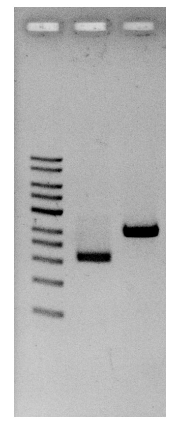
Plasmid DNA were loaded on 1% of agarose gel after extraction.
Comparison of workflows of Magnetic Beads based kits
Features
100% centrifuge-free
Culture medium can be used directly without centrifuge to pellet the bacteria
Simple
No centrifuge
No column
No vacuum
Flexible applications with simplified miniprep protocol
High throughput: 96-well plates with 0.2 ml of each culture (Cat.# 50011)
Low throughput: 1.5 ml tubes with 0.2 ml of each culture (Cat.# 50011)
Low throughput: 1.5 ml tubes with 2 ml of each culture (Cat.# 50012)
Our Blyscan™ Glycosaminoglycan Kit has been a ‘go-to’ Solution for reliable sGAG and Proteoglycan Analysis for many years! Blyscan utilises a dye-binding approach to quantitatively measure sulfated glycosaminoglycans (sGAG) and proteoglycans in cells, tissues and fluids from a wide range of in-vivo and in-vitro sources.
Colorimetric Detection (656nm) (Endpoint)
Understanding Glycosaminoglycans (GAGs) and Proteoglycans
Glycosaminoglycans (GAGs) are a type of negatively charged polysaccharide that play crucial roles in various biological processes. They are composed of repeated disaccharide units, typically of N-acetylated or N-sulfated hexosamine paired with a uronic acid (GlcA or IdoA) or galactose. Sulfate groups can also be added to give sulfated GAGs an overall negative charge that influences cell interactions and also enable binding by our Blyscan dye reagent.
Common examples of GAGs include Chondroitin Sulfate, Dermatan Sulfate, Heparin, Heparan Sulfate, and Keratan Sulfate. Note that Hyaluronic Acid is a non-sulfated GAG and cannot be detected by the Blyscan assay. If you need to measure hyaluronic acid instead, we recommend using our Purple-Jelley kit!
The Role of Glycosaminoglycans in Tissues
GAGs and proteoglycans have essential functions in tissues and organisms, providing biophysical support through scaffolding and maintaining cartilage hydration. They also play a vital role in biochemical processes such as cell adhesion and signalling.
What is the origin of the Blyscan assay name?
Blyscan is an Old English word meaning ‘to shine’ and from which the word ‘blush’, (blushing), may have been derived. This was an appropriate choice as the Blyscan Assay contains a blue dye which ‘blushes’ bright pink when it binds to sulphated glycosaminoglycans!
How does the Blyscan assay work?
Step 1. Blyscan dye reagent contains DMMB dye in an optimised buffer. Addition of Dye reagent to samples containing sGAG results in the formation of a dye/sGAG complex due to a charge interaction between dye and GAG sulfate groups.
Step 2. Over a 30 minute incubation Dye-labelled sGAGs precipitate out of solution and are collected by centrifugation. Following removal of unbound dye, the remaining bound dye is released from the complex by addition of dye dissociation reagent. Released dye is quantified spectrophotometrically.
Step 3. The sGAG content of unknown samples may be quantified by comparison against a calibration curve prepared using a standard of purified Chondroitin-4-sulfate supplied with the kit.
A list of suggested sample types can be found under the ‘Assay Specification‘ tab.
The Blyscan Dye reagent is formulated to miminise binding to other charged sample components such as nucleic acids, a problem with some older dye-based sGAG assays.
Assay range
2.5 – 50µg/ml
Limit of Detection
2.5µg/ml
Detection Method
Colorimetric Detection (656nm) (Endpoint)
Measurements per kit
110 in total (allows a maximum of 48 samples to be run in duplicate alongside a standard curve).
In-vivo: Liquid samples, including fluids such as urine, amniotic or synovial fluid.
In-vitro: Solid samples, such as deposited ECM on 2D/3D culture surfaces.by enzymatic treatment
In-vivo: Liquid samples, Culture media during 2D/3D cell culture.
The assay requires that sulfated polysaccahrides or sGAGs are in a soluble form. A preliminary enzymatic extraction step is required for solid samples (enzyme not supplied with kit).
The assay is not suitable for use with samples containing alginates or that comprise degraded sulfated disaccharide fragments.
Precautions
This kit is designed for research use only. Not for use in diagnostic procedures. Kit requires access to a centrifuge, as well as a spectrophotometer/colorimeter capable of absorbance detection at 656nm. Specific sample preparation protocols may require customer to provide further reagents, consult assay manual for further information.
Blyscan sGAG kit contents:
1. Blyscan Dye Reagent (1x110ml)
2.sGAG Reference Standard (1x5ml, 100µg/ml Bovine tracheal chondroitin 4-sulfate)
3. Dissociation Reagent (1x110ml)
4. Sodium Nitrite (1x15ml)
5. Acetic Acid (1x15ml)
6. Ammonium Sulfamate (1x15ml)
7. 1.5ml micro-centrifuge tubes for dye-labelling reaction.
8. Assay kit manual
NB: Additional reagents may be required for sample preparation prior to assay. Consult manual or contact us for further details.
Document
Our Blyscan™ Glycosaminoglycan Kit has been a ‘go-to’ Solution for reliable sGAG and Proteoglycan Analysis for many years! Blyscan utilises a dye-binding approach to quantitatively measure sulfated glycosaminoglycans (sGAG) and proteoglycans in cells, tissues and fluids from a wide range of in-vivo and in-vitro sources.
Colorimetric Detection (656nm) (Endpoin
This product provides a fast and easy way to purify DNA from plant and fungal tissue. Up to 3g of tissue can be processed. Easy-to-use Plant procedures provide pure total DNA (genomic, mitochondrial, and chloroplast) for reliable PCR and southern blot in less than 1 hour.
Details
Specifications
Features
Specifications
Main Functions
Isolation total DNA from 3g plant and fungal tissue
Applications
PCR, SSR, AFLP, RAPD and southern blot, etc.
Purification method
Mini spin column
Purification technology
Silica technology
Process method
Manual (centrifugation or vacuum)
Sample type
Various plant samples (including conventional, polysaccharides and polyphenols)
This product is based on silica Column purification. Plant material is first mechanically disrupted and then lysed by addition of lysis buffer and incubation. RNase A in the lysis buffer digests the RNA in the sample. After lysis, proteins and polysaccharides are salt-precipitated. Binding buffer and ethanol are added to the cleared lysate to promote binding of the DNA to the HiPure membrane. The sample is then applied to a column and then centrifuged.DNA binds to the membrane, while contaminants such as proteins and polysaccharides are efficiently removed by 2 wash steps. Pure DNA is eluted in a small volume of low-salt buffer or water.
Advantages
Broad spectrum – suitable for extracting DNA from various plant samples
Fast – several samples can be extracted in 30 minutes by silica technology
High purity – high quality DNA, completely remove inhibitors
High yield – silica technology can achieve the highest yield
Kit Contents
Contents
D316302
D316303
Purification Times
10 Preps
50 Preps
RNase A
20 mg
90 mg
Protease Dissolve Buffer
1.8 ml
10 ml
Buffer PAL
180 ml
900 ml
Buffer GWP
150 ml
800 ml
Buffer GW2
25 ml
200 ml
Buffer AE
30 ml
120 ml
HiPure DNA Maxi Columns II
10
50
50 ml Collection Tubes
20
100
Storage and Stability
RNase A should be stored at 2-8°C upon arrival. However, short-term storage (up to 12 weeks) at room temperature (15-25°C) does not affect its performance. The remaining kit components can be stored dry at room temperature (15-25°C) and are stable for at least 18 months under these conditions. The entire kit can be stored at 2-8°C, but in this case buffers should be redissolved before use. Make sure that all buffers are at room temperature when used.
This product provides a fast and easy way to purify DNA from plant and fungal tissue. Up to 3g of tissue can be processed. Easy-to-use Plant procedures provide pure total DNA (genomic, mitochondrial, and chloroplast) for reliable PCR and southern blot in less than 1 hour.
Permagen’s 6 x 1.5 mL Microfuge Tube Magnetic rack is designed for magnetic bead separations from up to 6 tubes
Accommodates many common 1.5 mL Microcentrifuge and some 2.0 mL tubes
Tubes are angled and beads will be pulled to back wall allowing easy aspiration and tip tracking down the front wall of the tubes without disturbing bead pellet