[PM1700] ExcelBand™ All Blue Broad Range Protein Marker (9-240 kDa), 250 μl x 2
Facebook
X
Pinterest
Email
Detail
Description
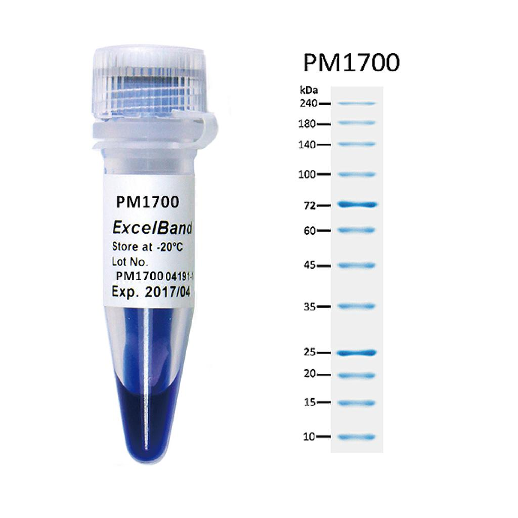
The PM1700 ExcelBand™ All Blue Broad Range Protein Marker is a blue protein standard with 12 pre-stained proteins covering a wide range of molecular weights from 10 to 240 kDa in Tris-Glycine buffer (9 to 235 kDa in Bis-Tris (MOPS) buffer and Bis-Tris (MES) buffer). Proteins are covalently coupled with a blue chromophore, and two reference bands (at 25 kDa and 72 kDa, respectively) are enhanced in intensity when separated on SDS-PAGE (Tris-Glycine buffer).
The PM1700 ExcelBand™ All Blue Broad Range Protein Marker is designed for monitoring protein separation during SDS-polyacrylamide gel electrophoresis, verification of Western transfer efficiency on membranes (PVDF, nylon, or nitrocellulose) and for approximating the size of proteins.
Features
Ready-to-use — Premixed with a loading buffer for direct loading, no need to boil.
Two enhanced bands — 72 kDa and 25 kDa
Contents
Approximately 0.1~0.5 mg/ml of each protein in the buffer (20 mM Tris-phosphate (pH 7.5 at 25°C), 2% SDS, 0.2 mM DTT, 3.6 M urea, and 15% (v/v) glycerol).
Quality Control
Under suggested conditions, PM1700 ExcelBand™ All Blue Broad Range Protein Marker resolves 12 major bands in SDS-PAGE (Tris-Glycine buffer, MOPS, and MES buffer) and after Western blotting to nitrocellulose membrane.
ChIP-Seq Library Prep Kit (illumina and MGI Platforms)
Product Info
Document
Product Info
The ChIP-Seq Library Prep Kit (illumina and MGI Platform) was developed for the construction of high quality ChIP-Seq libraries using 5 ng to 400 ng of ChIP DNA as input. The kit is compatible with ChIP DNA fragments generated from both enzymatic methods and physical methods (sonication, nebulization etc.).
ChIP-Seq Library Prep Kit Workflow
ChIP-Seq is the combination of chromatin immunoprecipitation (ChIP) with next generation sequencing. It is a powerful tool for the analysis of global transcription factors and other proteins in diseases and biological pathways, and characterization of histone modifications in a genome-wide level at single-base resolution. ChIP-Seq delivers whole genome level of functional profiling of global transcription factors, and provides better understanding of epigenetic modifications.
Three index types are available for the ChIP-Seq Library Prep Kit of the illumina platform:
Non-index (Cat.# 30032): Libraries do not have index.
Index (Cat.# 30034): Each index primer contains a unique 6-base index sequence can be used for identification. 48 samples can be pooled together. Index information can be downloaded here.
Unique dual index (Cat.# 30036): The ChIP-Seq library multiplexing for 96 samples is possible. Our unique 4-Base Difference Index System have 8 bases index length and at least 4 bases are different from each other for better library identification. Our unique dual indexing primers remove sequencing errors such as index hopping, index contamination, mis-assignment, and other errors. Index information can be downloaded here.
Indexes are available for the MGI platform kits (Cat.# 34034).
Kit advantages:
Super fast protocol
Library prep can be done in 1.5 hours
The hands-on time is only around 10 minutes
Easy procedure
Ready-to-use master mix simplified the procedure
Less reaction components make it easy to setup reactions
Reduced more than half of the beads cost
Input ChIP DNA: From 5 ng to 400 ng
Comparison of library conversion efficiency under the same condition. Input DNA amounts are 5 ng and 30 ng. BioDynami ChIP-Seq Library Prep Kit (Cat.# 30034) was used.
Comparison of aligned reads, aligned rate and duplication rate. Input DNA amounts are 5 ng and 30 ng. BioDynami ChIP-Seq Library Prep Kit (Cat.# 30034) was used.
Data comparison: Input DNA amounts are 5 ng and 30 ng. BioDynami ChIP-Seq Library Prep Kit (Cat.# 30034) was used. Sequencing peak regions are shown.
Document
The ChIP-Seq Library Prep Kit (illumina and MGI Platform) was developed for the construction of high quality ChIP-Seq libraries using 5 ng to 400 ng of ChIP DNA as input. The kit is compatible with ChIP DNA fragments generated from both enzymatic methods and physical methods (sonication, nebulization etc.).
The BK virus is a member of the polyomavirus family. BK viral infections are typically asymptomatic in healthy individuals, however very mild symptoms may appear including mild respiratory infections and fever. Once an individual has been infected the virus disseminates to the kidneys and the urinary tract where it remains for the lifetime of the individual. Infections with BK virus in immunocompromised or immunosupressed patients are much more severe and may involve renal dysfunction. In fact, in kidney transplant patients the immunosupressive drugs required for the transplant may allow the virus to replicate within the graft, resulting in a disease called BK virus nephropathy (BKVN). It is thought that 1-10% of renal transplant patients progress to BK virus nephropathy (BKVN) and up to 80% of these patients are reported to have lost their grafts. The onset of nephritis can occur as early as several days post-transplant to as late as 5 years. The mode of transmission of the virus is not clear, however it has been suggested that BKV may be transmitted through respiratory fluids or urine, since infected individuals periodically excrete virus in the urine. This virus can be diagnosed by BKV blood & urine testing, in addition to carrying out a biopsy in the kidneys. PCR techniques are now widely used to identify the virus.
BKV TaqMan PCR Kit, 100 reactions
Ready to use format, including Master Mix for the target and PCR control to monitor for PCR inhibition and validate the quality
Specific Primer and Probe mix for the pathogen/virus/viroid of interest
Primer and Probe mix
Positive and negative control to confirm the integrity of the kit reagents
BKV TaqMan PCR Probe/Primer Set and Controls, 100 reactions
Specific Primer/Probe mix and Positive Control for the pathogen/virus/viroid of interest
Nuclease-free water
Can be used together with Norgen’s PCR Master Mix (#28007) or customer supplied master mix
For research use only and NOT intended for in vitro diagnostics.
Storage Conditions and Product Stability All kit components can be stored for 2 years after the date of production without showing any reduction in performance.
All kit components should be stored at -20°C upon arrival.