

Description
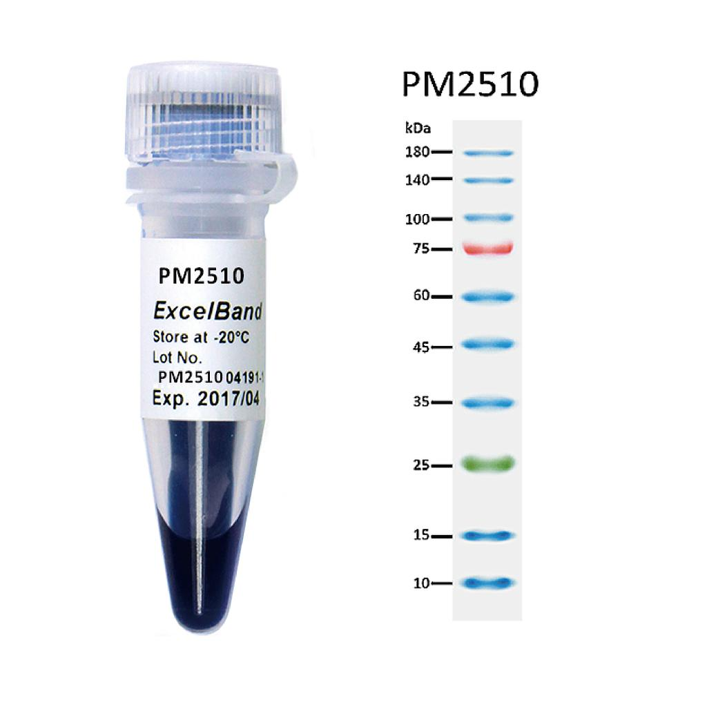
The PM2510 ExcelBand™ Enhanced 3-color Regular Range Protein Marker is a ready-to-use three-color protein standard with 10 pre-stained proteins covering a wide range of molecular weights from 10 to 180 kDa in Tris-Glycine Buffer (9 to 170 kDa in Bis-Tris (MOPS) buffer and 10 to 170 kDa Bis-Tris (MES) buffer). Proteins are covalently coupled with a blue chromophore except for two reference bands (one green and one red band at 25 kDa and 75 kDa respectively) when separated on SDS-PAGE (Tris-Glycine buffer). PM2510 ExcelBand™ Enhanced 3-color Regular Range Protein Marker is designed for monitoring protein separation during SDS-polyacrylamide gel electrophoresis, verification of Western transfer efficiency on membranes (PVDF, nylon, or nitrocellulose) and for approximating the size of proteins.
Features
- Ready-to-use — Premixed with a loading buffer for direct loading, no need to boil.
- Two reference bands — 75 kDa (red) and 25 kDa (green)
Contents
Approximately 0.2~0.6 mg/ml of each protein in the buffer (20 mM Tris-phosphate (pH 7.5), 2% SDS, 3.6 M urea, and 15% (v/v) glycerol).
Quality Control
Under suggested conditions, PM2510 ExcelBand™ Enhanced 3-color Regular Range Protein Marker resolves 10 major bands in 15% SDS-PAGE (Tris-Glycine buffer, MOPS, and MES buffer) and after Western blotting to nitrocellulose membrane.
Storage
4°C for 3 months
-20°C for long term storage


