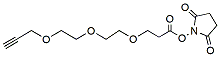
Propargyl-PEG3-NHS ester is a Click Chemistry reagent with a propargyl group and an NHS ester group. The propargyl group can react with biomolecules containing azide group via copper catalyzed Click Chemistry reaction. The NHS ester is an amine reactive group which can be used for derivatizing peptides, antibodies, amine coated surfaces etc. Reagent grade, for research purpose. Please contact us for GMP-grade inquiries.
Detail
Propargyl-PEG3-NHS ester is a Click Chemistry reagent with a propargyl group and an NHS ester group. The propargyl group can react with biomolecules containing azide group via copper catalyzed Click Chemistry reaction. The NHS ester is an amine reactive group which can be used for derivatizing peptides, antibodies, amine coated surfaces etc. Reagent grade, for research purpose. Please contact us for GMP-grade inquiries.
Other Products
NGS Library Circularization Kit (MGI Platform)
Product Info
Document
Product Info
The NGS Library Circularization Kit (MGI Platform) was developed for preparation of single-stranded circular DNA libraries for next generation sequencing (MGI platform).
The kit uses linear dsDNA libraries (MGI platform) as input and enriches the circularized single-stranded DNA libraries. The circularization kit has a higher library circularization efficiency (25%) as compared to other vendors (7-15%).
NGS Library Circularization Kit workflow
Comparison of Library Circularization Efficiency
Kit features
Fast protocol
Total protocol time is around 1 hour
Hands-on time is only ~10 minutes
Guaranteed high library circularization efficiency as compared to other kits
Input DNA amount: 100-300 ng
Document
The NGS Library Circularization Kit (MGI Platform) was developed for preparation of single-stranded circular DNA libraries for next generation sequencing (MGI platform).
【IT1000】EzRNA™ T7 High Yield RNA Synthesis Kit, 50 RXN
Product Info
Document
Product Info
Description
The EzRNA™ T7 High Yield RNA Synthesis Kit is a user-friendly product for enzymatic RNA production. The enzyme mix contains adequate amount of T7 RNA polymerase, pyrophosphatase, and RNase inhibitors for in vitro transcription (IVT). Along with 10X Transcription Buffer and NTP Premix, users can swiftly assemble IVT reactions without compromising RNA yield. The EzRNA™ T7 High Yield RNA Synthesis Kit allows for the attainment of approximately up to 150 µg RNA yield within 2 hours at 37°C.
Features
High yield
Versatile- suitable for short and long transcripts
NTP premixed- Minimal pipetting and setup time
Compatible with CleanCap® Reagent AG
Lithium chloride included for RNA purification
Application
Generation of RNA from T7 promoter-driven DNA sequences
Suitable for subsequent cap-0 and cap-1 modification
Storage
-20°C for 12 months
Document
The EzRNA™ T7 High Yield RNA Synthesis Kit is a user-friendly product for enzymatic RNA production. The enzyme mix contains adequate amount of T7 RNA polymerase, pyrophosphatase, and RNase inhibitors for in vitro transcription (IVT). Along with 10X Transcription Buffer and NTP Premix, users can swiftly assemble IVT reactions without compromising RNA yield. The EzRNA™ T7 High Yield RNA Synthesis Kit allows for the attainment of approximately up to 150 µg RNA yield within 2 hours at 37°C.
The BK virus is a member of the polyomavirus family. It has been suggested that this virus may be transmitted through respiratory fluids or urine, since infected individuals periodically excrete virus in the urine. BK viral infections are typically asymptomatic in healthy individuals, however very mild symptoms may appear including mild respiratory infections and fever. Infections with BK virus in immunocompromised or immunosupressed patients are much more severe and may involve renal dysfunction. In fact, in kidney transplant patients the immunosupressive drugs required for the transplant may allow the virus to replicate within the graft, resulting in a disease called BK virus nephropathy (BKVN). The JC virus is a type of human polyomavirus and is very common in the general population, infecting 70 to 90% of humans. Most people acquire JCV in childhood or adolescence. Typically the infection is subclinical and no of consequence in individuals with healthy immune systems. The initial site of infection may be the tonsils or the gastrointestinal tract, and the virus then remains latent in the gastrointestinal tract. JCV can also infect the tubular epithelial cells in the kidneys, where it continues to reproduce, shedding virus particles in the urine. Also, JCV can cross the blood-brain barrier into the central nervous system. JCV is known to cause the usually fatal progressive multifocal leukoencephalopathy (PML) by destroying oligodendrocytes in the brain in immunodeficient or immunosuppressed individuals. The JC and BK viruses are very similar, with their genomes sharing 75% homology. It is however important to differentiate between the viruses due to the differences in pathology and especially the invariably fatal outcome of PML which is only caused by the JC virus.
Storage Conditions and Product Stability All kit components can be stored for 2 years after the date of production without showing any reduction in performance.
All kit components should be stored at -20°C upon arrival.