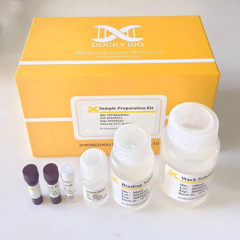
Note: Price not include shipment & duty, contact us to get full quote. The Residual DNA Sample Preparation Kit uses chemical lysis and magnetic beads to extract DNA from diverse sample types, including samples that contain high protein and low DNA concentration. The kit extracts residual genomic DNA from products that are produced in cell lines. The operation is easier and the higher nucleic acid yield is guaranteed at the same time.
Detail
Description
The Residual DNA Sample Preparation Kit uses chemical lysis and magnetic beads to extract DNA from diverse sample types, including samples that contain high protein and low DNA concentration. The kit extracts residual genomic DNA from products that are produced in cell lines such as CHO, E. coli, E1A, HEK293, Vero, NS0 and Baculovirus. For quantification of residual DNA, we recommend using the resDNASEQ Residual DNA Quantitation kit as described in the resDNASEQ Residual DNA Quantitation kit User Guide. To ensure accurate quantitative results, each sample in triplicate and perform a single PCR reaction for each extraction.
Video Player
Features of theresDNASEQ E.coli Residual DNA Quantitation kit include:
Simpler and Rapid
Only three steps will be need for Sample Preparation,and the total time of Sample preparation is about 30 minites.
Good Recovery
The good Recovery rate of Sample Preparation.
Easy to store
All components of the Sample Preparation Kit can be stored at room temperature.
Other Products
D2111 HiPure Gel DNA Mini Kit
Product Info
Document
Product Info
Introduction
HiPure Gel Pure DNA Kit uses proprietary chemistry and HiPure technology to recover DNA fragments between 60bp-10kbp with yields exceeding 80%. DNA is suitable for ligations, PCR, sequencing, restriction digestion, or various labeling reactions. In addition, this kit can be also used to recover DNA directly from enzymatic reactions such as PCR and enzyme digestion reactions.
Details
Specifications
Features
Specifications
Main Functions
Recover DNA fragments >100bp from agarose gel(<0.5g), purification of DNA from PCR, enzymatic reaction solution or crude gDNA
Applications
PCR, NGS, labeling, ligation and enzyme digestion, etc.
Purification method
Mini spin column
Purification technology
Silica technology
Process method
Manual (centrifugation or vacuum)
Sample type
Agarose gel, PCR products, enzyme products
Sample amount
Agarose gel: ≤500mg
Recovery
≥80%
Elution volume
≥15μl
Time per run
≤20 minutes(1-24 samples)
Liquid carrying volume per column
800µl
Binding yield of column
35µg
Principle
The HiPure system uses a simple bind-wash-elute procedure. Gel slices are dissolved in a buffer containing a pH indicator, allowing easy determination of the optimal pH for DNA binding, and the mixture is applied to the column. Nucleic acids adsorb to the silica-gel membrane in the high-salt conditions provided by the buffer. Impurities are washed away and pure DNA is eluted with a small volume of low-salt buffer provided or water, ready to use in subsequent applications.
Advantages
High recovery efficiency – ≥80% DNA recovery
General – recover DNA from gel or enzyme-driven reaction solutions such as PCR
Fast – isolation can be completed in 10-15 minutes by column gel method
Great cost-effectiveness performance
Kit Contents
Contents
D211102
D211103
Purification Times
100 Preps
250 Preps
Buffer GDP
120 ml
250 ml
Buffer DW2
50 ml
2 x 50 ml
Elution Buffer
20 ml
30 ml
HiPure DNA Mini Columns II
100
250
2 ml Collection Tubes
100
250
Storage and Stability
The Kit should be stored dry at room temperature (15-25°C) and are stable for at least 18 months under these conditions. If any precipitates form in the buffers, warm at 37℃ to dissolve.
Experiment Data
Document
HiPure Gel Pure DNA Kit uses proprietary chemistry and HiPure technology to recover DNA fragments between 60bp-10kbp with yields exceeding 80%. DNA is suitable for ligations, PCR, sequencing, restriction digestion, or various labeling reactions. In addition, this kit can be also used to recover DNA directly from enzymatic reactions such as PCR and enzyme digestion reactions.
How to use: 1.Suspend 18g in 1L of distilled water, stirring heated to boiling to completely dissolve, autoclave at 121℃ for 15 minutes. 2.Diluted and treated samples.
Quality control:
Item
The name and number of strain
Growth
Colony Color
1
Escherichia coli ATCC25922
Good
Turbid broth,slight precipitate
2
Staphylococcus aureus ATCC25922
Good
Turbid broth,slight precipitate
Storage: Keep container tightly closed, store in a cool, dry place, away from bright light. Storage period of 3 years.
Short term stability: 2-8oC, Long term stability: See individual component labels
Stability:
> 2 years under recommended storage conditions
Analyte:
D-Fructose, D-Glucose, D-Mannose
Assay Format:
Spectrophotometer
Detection Method:
Absorbance
Wavelength (nm):
340
Signal Response:
Increase
Linear Range:
4 to 80 µg of D-glucose, D-fructose or D-mannose per assay
Limit of Detection:
~ 0.7 mg/L
Reaction Time (min):
~ 30 min
Application examples:
Foodstuffs, yeast cell preparations, enzymatic hydrolysates and other materials (e.g. biological cultures, samples, etc.).
Method recognition:
Novel method
The D-Mannose/D-Fructose/D-Glucose test kit is suitable for the specific measurement and analysis of D-mannose, D-fructose and D-glucose in plant products and in acid hydrolysates of polysaccharides.
Note for Content: The number of manual tests per kit can be doubled if all volumes are halved. This can be readily accommodated using the MegaQuantTM Wave Spectrophotometer (D-MQWAVE).
All reagents stable for > 2 years after preparation
Only enzymatic kit available
Simple format
Rapid reaction
Mega-Calc™ software tool is available from our website for hassle-free raw data processing
Standard included
Document
The D-Mannose/D-Fructose/D-Glucose test kit is suitable for the specific measurement and analysis of D-mannose, D-fructose and D-glucose in plant products and in acid hydrolysates of polysaccharides.