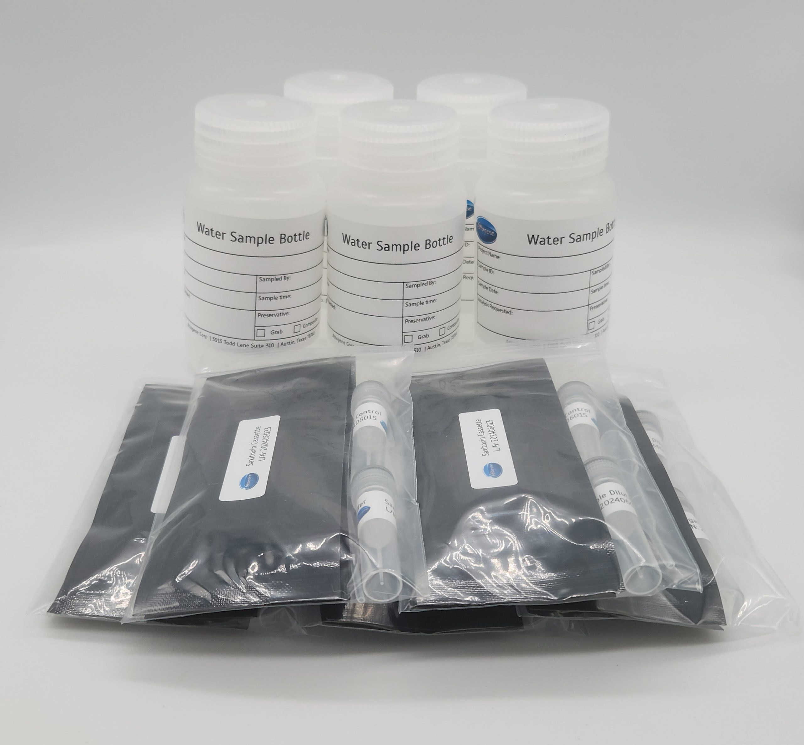
Saxitoxin (PSP) Lateral Flow Kit (Freshwater Streams and Source Water)
Facebook
X
Pinterest
Email
Screening of saxitoxin in Freshwater Streams and Source Water samples as low as 70 ppt Format: 10 tests (5 tests/5 control) Water Sample Bottles provided Run Time: 15 Minutes
Detail
Screening of saxitoxin in Freshwater Streams and Source Water samples as low as 70 ppt
Saxitoxins (STXs) are naturally occurring alkaloids produced by some marine dinoflagellates and by strains of various species of freshwater cyanobacteria. Saxitoxin is one of the prevalent paralytic shellfish toxins (PSTs). It belongs to a family of potent neurotoxins with a molecular weight around 300 Da. Saxitoxin and its derivatives are alkaloids composed of a tetrahydropurine ring system with a highly polar guanidinium group. Due to their significant toxicity, saxitoxins are closely monitored in marine environments where they can accumulate during harmful algal blooms (HABs). In 2009 an action level of 0.6ppb (600ppt) was recommended by EFSA as a guidance framework have been established to manage saxitoxin levels in water.
Q-PAGE™ Bis-Tris Precast Gel is a high-performance and easy to use precast polyacrylamide gel for electrophoresis in Bis-Tris buffer system (MOPS or MES). The optimized gel formula allows Q-PAGE™ Bis-Tris Precast Gel to show improved resolution, accurate results, and an extended shelf-life over conventional Laemmli Tris-HCl gels.
Q-PAGE™ Bis-Tris Precast Gels are available in gradient (4 to 12%) and fixed (8% and 12%) concentrations of polyacrylamide in 12-and 15-well formats. Two available cassette sizes, Mini (10 x 8.3 cm) and Midi (10 x 10 cm), are compatible with most popular protein electrophoresis systems. Q-PAGE™ Mini (QP2XXX) Gels are suitable for Bio-Rad® and other systems. Q-PAGE™ Midi (QP3XXX) Gels are suitable for Invitrogen® XCell SureLock® Mini-Cell, Invitrogen® Mini Gel Tank, Hoefer SE260, and other systems.
Key Features
User-friendly gel cassette:
Numbered and framed wells for sample loading
With cassette opener for easy use
Enhanced gel performance:
Enhanced band sharpness
Better resolution of small proteins
Stable for shipping at ambient temperature
Easy compatibility:
Available as homogeneous and adjusted gradient gels for a wide range of protein separation.
Compatible with most popular protein electrophoresis systems
Storage and stability
Store Q-PAGE™ Precast Gels at 4°C for periods up to 12 months.
Do not freeze Q-PAGE™ Precast Gels Remove tape and comb before electrophoresis.
Keep Q-PAGE™ Precast Gels flat during storage.
Document
Q-PAGE™ Bis-Tris Precast Gel is a high-performance and easy to use precast polyacrylamide gel for electrophoresis in Bis-Tris buffer system (MOPS or MES). The optimized gel formula allows Q-PAGE™ Bis-Tris Precast Gel to show improved resolution, accurate results, and an extended shelf-life over conventional Laemmli Tris-HCl gels.
Q-PAGE™ Bis-Tris Precast Gels are available in gradient (4 to 12%) and fixed (8% and 12%) concentrations of polyacrylamide in 12-and 15-well formats. Two available cassette sizes, Mini (10 x 8.3 cm) and Midi (10 x 10 cm), are compatible with most popular protein electrophoresis systems. Q-PAGE™ Mini (QP2XXX) Gels are suitable for Bio-Rad® and other systems. Q-PAGE™ Midi (QP3XXX) Gels are suitable for Invitrogen® XCell SureLock® Mini-Cell, Invitrogen® Mini Gel Tank, Hoefer SE260, and other systems.
Bis-Sulfone-PEG9-DBCO is a bis-alkylating labeling reagent that is selective for the cysteine sulfur atoms from a native disulfide. These reagents undergo bis-alkylation to conjugate both thiols derived from the two cysteine residues of a reduced native disulfide bond such as the interchain disulfide bonds of an antibody. The reaction results in covalent rebridging of the disulfide bond via a three carbon bridge leaving the protein structurally intact. Reagent grade, for research purpose. Please contact us for GMP-grade inquiries.
Document
Bis-Sulfone-PEG9-DBCO is a bis-alkylating labeling reagent that is selective for the cysteine sulfur atoms from a native disulfide. These reagents undergo bis-alkylation to conjugate both thiols derived from the two cysteine residues of a reduced native disulfide bond such as the interchain disulfide bonds of an antibody. The reaction results in covalent rebridging of the disulfide bond via a three carbon bridge leaving the protein structurally intact. Reagent grade, for research purpose. Please contact us for GMP-grade inquiries.
Hipure Stool RNA Kit is specially designed for stool RNA extraction. This kit is suitable for extracting high-purity microbial or host cell RNA from ≤0.1g stool samples. The kit adopts silica gel column purification technology and original solution system, which can effectively remove humic acid and other inhibitory factors in stool samples. The purified RNA can be directly used in RT-PCR, Northern hybridization and other experiments.
Details
Specifications
Features
Specifications
Main Functions
Isolation total RNA from 100-150mg stool sample
Applications
RT-PCR, Northern hybridization and other experiments
Purification method
Mini spin column
Purification technology
Silica technology
Process method
Manual (centrifugation or vacuum)
Sample type
Stool
Sample amount
100-150 mg
Elution volume
≥30μl
Time per run
≤50 minutes
Liquid carrying volume per column
100µg
Binding yield of column
800µl
Principle
The HiPure silica gel column uses a high binding ability glass fiber filter membrane as the substrate. Under the condition of high concentration of ionizing agent (such as Guanidinium chloride or guanidine isothiocyanate), the filter membrane can adsorb nucleic acid through hydrogen bonding and electrostatic and other physical factors, while protein or other impurities are not adsorbed and removed. The filter membrane that has adsorbed nucleic acids is washed to remove proteins and salts. Finally, low salt buffer solution (such as Buffer TE) or water can be used to wash out the nucleic acids adsorbed on the filter membrane. The obtained nucleic acid has high purity and can be directly used in various downstream experiments.
The stool samples are homogenized in the lysis solution, further lysed in a high-temperature water bath, and RNA is released into the lysis solution. Chloroform extraction removes genomic DNA and impurities, transfer the supernatant to an alcohol free binding solution, purify RNA through a column, and finally elute RNA with RNase Free Water. The purified RNA can be directly used for experiments such as PCR, Southern hybridization, and enzyme digestion.
Advantages
High purity – unique adsorbent for more efficient removal of inhibitors
High concentration – maximum extraction of total RNA from stool samples
Sensitive – RNA can be purified at the level of PG
Kit Contents
Contents
R418502
R418503
Purification Times
50 Preps
250 Preps
HiPure RNA Mini Columns
50
250
2ml Collection Tubes
50
250
Glass Beads (0.1~0.6mm)
30 g
150 g
Buffer SPL
30 ml
140 ml
Buffer PHC
30 ml
140 ml
Buffer GRP
60 ml
250 ml
Buffer RW1
50 ml
250 ml
Buffer RW2 *
20 ml
2 x 50 ml
RNase Free Water
15 ml
30 ml
Storage and Stability
The kit components can be stored at room temperature (15–25°C) and are stable for 18 months under these conditions. At low temperatures, Buffer SPL may form precipitates, dissolve it by 55°C water bath. After receiving the product, Buffer PHC should be stored at 2-8°C.
Document
Hipure Stool RNA Kit is specially designed for stool RNA extraction. This kit is suitable for extracting high-purity microbial or host cell RNA from ≤0.1g stool samples. The kit adopts silica gel column purification technology and original solution system, which can effectively remove humic acid and other inhibitory factors in stool samples. The purified RNA can be directly used in RT-PCR, Northern hybridization and other experiments.