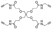
Tetra(3-methoxy-N-(prop-2-ynyl)propanamide) Methane is a 4-branched molecule with propargyl groups that can be linked to azide-containing biomolecules via Click Chemistry. Reagent grade, for research purpose. Please contact us for GMP-grade inquiries.
Detail
Tetra(3-methoxy-N-(prop-2-ynyl)propanamide) Methane is a 4-branched molecule with propargyl groups that can be linked to azide-containing biomolecules via Click Chemistry. Reagent grade, for research purpose. Please contact us for GMP-grade inquiries.
Other Products
FFPE RNA Purification Kits
Product Info
Document
Product Info
Overview
Extract total RNA (including microRNA) from FFPE samples
No phenol extraction step
Includes DNase for optional on-column DNA removal
Isolated RNA is of the highest quality and integrity
Isolate a diversity of RNA species
Purified RNA is suitable for a variety of downstream applications, including Small RNA Sequencing. Find out more information on Norgen’s NGS services
Purification is based on spin column chromatography that uses Norgen’s proprietary resin separation matrix
Norgen’s FFPE RNA Purification Kits provide a rapid method for the isolation and purification of total RNA (including microRNA) from formalin-fixed paraffin-embedded (FFPE) tissue samples in as little as 1 hour. Using formalin to fix tissues leads to crosslinking of the RNA and proteins, and the process of embedding the tissue samples can also lead to fragmentation of the RNA over time. Norgen’s FFPE RNA Purification Kits provide conditions that allow for the partial reversing of the formalin modifications, resulting in a high quality and yield of RNA. These kits are able to purify all sizes of RNA, from large mRNA and ribosomal RNA down to microRNA (miRNA) and small interfering RNA (siRNA), depending on the age of the FFPE tissue as fragmentation of the RNA is known to occur over time. The RNA is preferentially purified from other cellular components without the use of phenol or chloroform.
FFPE RNA Purification Kit (Spin Column)
Maximum loading volume of 650 μL per column, and a maximum binding capacity of 50 μg per column.
Purification is based on 96-well column chromatography using Norgen’s proprietary resin as the separation matrix. Purification can be performed using either a vacuum manifold or centrifugation. Maximum loading volume of 400 μL per well, and a maximum binding capacity of 50 μg per well.
5 slices of < 20 µm thick paraffin slices 25 mg of unsectioned block
Average Yield
Variable due to age of paraffin blocks ~2-3 µg of Total RNA per 1 mg of fresh FFPE hamster kidney
* Time required for purification varies by length of Proteinase K incubation and formalin crosslink-reversal
Storage Conditions and Product Stability All solutions should be kept tightly sealed and stored at room temperature. The DNAse I and Proteinase K should be stored at -20°C upon arrival. This kit is stable for 1 year from the date of shipment.
HiPure FFPE RNA Kit supplies a simple and rapid RNA extraction for Formalin-fixed, paraffin-embedded (FFPE) tissue and sections samples. This kit is based on silica gel column purification technology, no phenol-chloroform extraction or alcohol precipitation. The whole extraction only takes 30 minutes (not including digestion time). RNA can be directly used for downstream applications such as RT-PCR, Northern blot, vitro translation and other experiments.
Details
Specifications
Features
Specifications
Main Functions
Isolation total RNA from FFPE tissue and section samples (with DNase)
Applications
RT-PCR, quantitative RT-PCR, Northern hybridization, Poly A purification, nucleic acid protection and in vitro translation
Purification method
Mini spin column
Purification technology
Silica technology, DNase
Process method
Manual (centrifugation or vacuum)
Sample type
FFPE tissue sample
Sample amount
6mg
Yield
20μg
Elution volume
≥10μl
Time per run
≤60 minutes
Liquid carrying volume per column
800µl
Binding yield of column
100µg
Principle
This product is based on silica column purification. Remove paraffin by Buffer DPS. Sample lysis with proteinase K digestion requires only 15 minutes. After lysis, samples are incubated at 80ºC for 15 minutes. Transfer to an adsorption column and RNA is adsorbed on the membrane, while protein is not adsorbed and is removed with filtration. After washing proteins and other impurities, RNA was finally eluted with low-salt buffer.
Advantages
High quality – high purity total RNA can be directly used in various sensitive downstream applications
Fast – several samples can be extracted in 60 minutes by column method
Safe – no phenol chloroform extraction required
Sensitive – RNA can be recovered at the level of PG
Efficient DNA removal – unique method to effectively remove genomic DNA
Kit Contents
Contents
R414402
D414403
Purification Times
50 Preps
250 Preps
HiPure RNA Micro Columns
50
250
2ml Collection Tubes
50
250
Buffer DPS
60 ml
250 ml
Buffer FRL
15 ml
60 ml
Buffer RLC
15 ml
60 ml
Buffer RWC*
10 ml
50 ml
Buffer RW2*
20 ml
2 x 50 ml
DNase I
600 µl
5 x 600 µl
DNase Booster Buffer
1.5 ml
6 ml
Protease Dissolve Buffer
1.8 ml
10 ml
Proteinase K
24 mg
120 mg
RNase Free Water
10 ml
20 ml
Storage and Stability
Proteinase K should be stored at 2–8°C upon arrival. DNase I should be stored at -20°C. However, short-term storage (DNase I up to 1 weeks, Proteinase K up to 8 weeks) at room temperature (15–25°C) does not affect their performance. The remaining kit components can be stored at room temperature (15–25°C) and are stable for at least 18 months under these conditions.
Experiment Data
Document
HiPure FFPE RNA Kit supplies a simple and rapid RNA extraction for Formalin-fixed, paraffin-embedded (FFPE) tissue and sections samples. This kit is based on silica gel column purification technology, no phenol-chloroform extraction or alcohol precipitation. The whole extraction only takes 30 minutes (not including digestion time). RNA can be directly used for downstream applications such as RT-PCR, Northern blot, vitro translation and other experiments.
RNA/microRNA/DNA/Proteins are preserved for more than 2 years at room temperature in Norgen’s Urine Preservative
Compatible with most DNA, Total RNA, microRNA and protein isolation methods
Preservative is available in a single dose liquid format (ampule)
Preservative is also available in a dried format in tubes – Urine Collection and Preservation Tubes
Urine Collection and Preservation Devices are available in 4 convenient sizes: 5 cc tubes, 15 cc tubes, 50 cc tubes, and 120 cc cups
Norgen’s Urine Preservative is designed for the rapid preservation of DNA, RNA, microRNA and proteins from fresh urine samples. In addition, the Urine Preservative eliminates the need to immediately process or freeze samples and allows the samples to be shipped to centralized testing facilities at ambient temperatures. The components of the Urine Preservative allow samples to be stored for over 2 years at room temperature with no detected degradation of urine DNA, RNA or proteins.
Norgen’s Urine Preservative is available in 2 convenient formats:
1. Urine Preservative Single Dose Ampules With this product Norgen’s Urine Preservative is supplied in a liquid format in single dose ampules. The user simply collects 5 – 50 mL of urine into a urine collection container and adds the contents of the Urine Preservative Single Dose (Cat# 18126). The urine and preservative are then mixed, and the urine nucleic acids and proteins are preserved at room temperature.
2. Urine Collection and Preservation Tubes Norgen’s Urine Preservative is also available in a dried format in Norgen’s Urine Collection and Preservation Tubes. The user simply collects urine into the tubes and mixes gently until the orange preservative pellet in the tube has dissolved. Norgen’s Urine Collection and Preservation Tubes are available in 3 convenient sizes:
Select Urine Tested with the Norgen Urine Collection and Preservation Tubes
Human Mouse Lynx Wolf Urine collected from snow
Available Sizes and Formats
Urine Collection and Preservation Tubes (15 cc) – Urine inputs from 5 – 15 mL
Storage Conditions and Product Stability All tubes should be kept tightly sealed and stored at room temperature (15 – 25°C) for up to 2 years without any reduction in performance.