
Description
Ultrapure dCTP supplied as sodium salt in purified water (pH 8.5).
Features
- Ideal for PCR amplification and cDNA synthesis
- Nuclease and ribonuclease free
Applications
- PCR
- RT-PCR
Storage
-20°C for 36 months

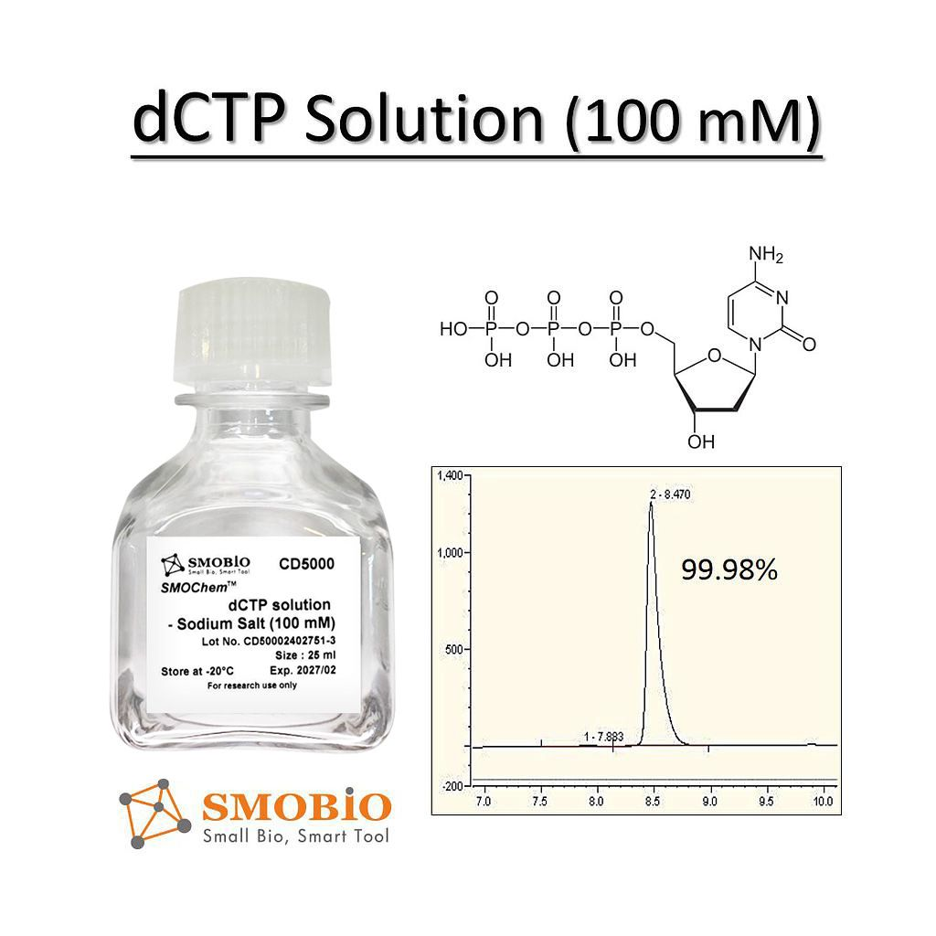
Description
Ultrapure dCTP supplied as sodium salt in purified water (pH 8.5).
Features
Applications
Storage
-20°C for 36 months

This kit provides a rapid spin column procedure for the purification and clean-up of sequencing and various other enzymatic reactions including restriction enzyme digests, Klenow reactions, alkaline phosphatase reactions, and ligations. The kit is used to remove reaction contaminants including dye terminators, salts, enzymes, excess primers and primer dimers. Contaminants are undesirable as they can interfere with many downstream applications including sequencing, RFLP, restriction enzyme digestions and ligation. Purification is based on spin-column chromatography without the use of phenol, chloroform or alcohol precipitation. The kit provides a high quality product with up to 90% recovery.
The kit is also available in a 96-well format for high-throughput sequencing reaction clean-up. Purification with the 96-well plate can be performed using either a vacuum manifold or centrifugation.
Figure 1 / 1

Click for expanded view
| Kit Specifications – 96-well | |
| Binding Capacity Per Well | 15 μg |
| Maximum Loading Volume Per Well | 500 μL |
| Average DNA Recovery | Up to 90% |
| Time to Complete 96 Purifications | 20 minutes |
| Minimum Elution Volume | 50 μL |
Storage Conditions and Product Stability
All solutions should be kept tightly sealed and stored at room temperature. This kit is stable for 1 year after the date of shipment.
| Component | Cat. 34500 (50 preps) | Cat. 34400 (2 x 96-well plate) |
|---|---|---|
| Binding Buffer C | 30 mL | 3 x 30 mL |
| Wash Solution A | 12 mL | 2 x 20 mL |
| Elution Buffer B | 8 mL | 2 x 15 mL |
| Spin Columns | 50 | – |
| Collection Tubes | 50 | – |
| Elution Tubes (1.7 mL) | 50 | – |
| 96-Well Plate | – | 2 |
| Adhesive Tape | – | 4 |
| 96-Well Collection Plate | – | 2 |
| 96-Well Elution Plate | – | 2 |
| Product Insert | 1 | 1 |

Attogene’s Human IgG/IgM universal fluorescent lateral flow assay kit is a ready-to-use, universal test strip, which is based on the lateral flow technology that uses 655nm Emission Quantum Dot particles containing streptavidin to conveniently capture biotinylated antigens. The device is designed to easily develop qualitative or quantitative rapid test systems for detection of anti-human IgG and IgM antibody that react to the any antigen that can be biotinylated (i.e. viral antigen, autoimmune antigen) and is easily customizable providing every laboratory with the possibility to perform assay feasibility.
Formats (fluorescent broad range UV light excitation range of 300nm to 400nm, 610nm emission) Streptavidin conjugate pad):
Antibody tests are a method of choice to determine if a person has been exposed to a pathogen or not. They are also incredibly valuable in the detection of autoantibodies that can be found in human autoimmune disorders. In this test, a biotinylated antigen (User supplied) is mixed with a biotinylated rabbit IgG (bind to goat anti rabbit control line) and sample (human sera or plasma) is simply mixed into with the specially designed assay running buffer in a well of the supplied 96-well plate, mixed and is then added to the sample port of the cassette. Generally, the reaction is complete in 10-15 minutes. It is very important to note that the relative stoichiometry between the biotinylated antigen, biotinylated rabbit IgG added, and the streptavidin gold is critical for assay optimization. The appropriate concentration of biotinylated antigen to use with strips is dependent upon the purity and sequence and a standard curve can be used to determine the relative ratio (generally between 1ng-100ng per test). A positive control line (biotin-rabbit IgG) antibody will bind to the goat anti rabbit (GAR) line on the test to ensure the assay is running appropriately.

K-AVCHO
SKU: 700004267
100 Assays of each per kit
| Content: | 100 assays of each per kit |
| Shipping Temperature: | Ambient |
| Storage Temperature: | Short term stability: 2-8oC, Long term stability: See individual component labels |
| Stability: | > 2 years under recommended storage conditions |
| Analyte: | Available Carbohydrates, Dietary Fiber |
| Assay Format: | Spectrophotometer |
| Detection Method: | Absorbance |
| Wavelength (nm): | 340 |
| Signal Response: | Increase |
| Linear Range: | 4 to 80 μg of D-glucose, D-fructose or D-galactose per assay |
| Limit of Detection: | 1.475 g/100 g |
| Reaction Time (min): | ~ 5 h |
| Application examples: | Food ingredients, food products and other materials. |
| Method recognition: | AOAC Method 2020.07 |
The Available Carbohydrates Assay Kit method is suitable for the determination of available carbohydrates (AVCHO) comprising *total digestible starch (TDS) plus maltodextrins, sucrose, D-glucose, D-fructose and lactose. New Improved method receiving ‘First Action’ status: AOAC 2020.07. This method is designed to simulate in vivo conditions in the human small intestine (i.e. a 4 h incubation time with PAA + AMG) in parallel with recent advances in Dietary Fiber (DF) methodology (K-RINTDF: AOAC Method 2017.16) and in accordance with the new (physiological based) definition of DF announced by Codex Alimentarius in 2009. Also, sucrose is hydrolysed with a specific “sucrase” enzyme which (unlike invertase which has been used traditionally for this reaction) has no action on fructo-oligosaccharides (FOS).
* Total digestible starch (TDS) is defined as starch that is digested in a 4 h period and is part of the carbohydrate that is available for digestion and absorption in the human small intestine.
See our full range of dietary fiber assay kits.
The Available Carbohydrates Assay Kit method is suitable for the determination of available carbohydrates (AVCHO) comprising *total digestible starch (TDS) plus maltodextrins, sucrose, D-glucose, D-fructose and lactose. New Improved method receiving ‘First Action’ status: AOAC 2020.07. This method is designed to simulate in vivo conditions in the human small intestine (i.e. a 4 h incubation time with PAA + AMG) in parallel with recent advances in Dietary Fiber (DF) methodology (K-RINTDF: AOAC Method 2017.16) and in accordance with the new (physiological based) definition of DF announced by Codex Alimentarius in 2009. Also, sucrose is hydrolysed with a specific “sucrase” enzyme which (unlike invertase which has been used traditionally for this reaction) has no action on fructo-oligosaccharides (FOS).