This kit is used for extracting total viral nucleic acid from non-cell/low cell content biological samples such as body fluid, serum, plasma, immersion solution, tissue homogenate supernatant, culture supernatant, etc., the extracted products can be used for clinical in vitro detection.
Detail
Introduction
This kit is used for extracting total viral nucleic acid from non-cell/low cell content biological samples such as body fluid, serum, plasma, immersion solution, tissue homogenate supernatant, culture supernatant, etc., the extracted products can be used for clinical in vitro detection.
Details
Specifications
Features
Specifications
Main Functions
Rapidly Extract viral DNA/RNA from 200μl non-cell/low cell content biological samples such as body fluid, serum, plasma, urine, immersion solution, tissue homogenate supernatant, etc.
This product is based on silica gel purification. The sample is lysed and digested with lysate and protease, DNA/RNA is released into the lysate. Transfer to an adsorption plate and filter column. DNA/RNA is adsorbed on the membrane, while protein is not adsorbed and is removed with filtration. After washing proteins and other impurities, DNA / RNA was finally eluted with low-salt buffer (10 MmTris, pH 8.0).
Advantages
Fast – column purification without PK digestion
High quality – high purity total RNA can be directly used in various sensitive downstream applications
Safe – no phenol chloroform extraction required
Sensitive – DNA/RNA can be recovered at the level of PG
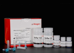
Kit Contents
Contents
IVD4175
Purification Times
100 Preps
HiPure Viral Column
100
2ml Collection Tubes
100
PK/Carrier RNA
50 mg/310μg
Protease Dissolve Buffer
5 ml
Buffer VLE
42 ml
Buffer CE
60 ml
RNase Free Water
15 ml
Storage and Stability
Proteinase K should be stored at 2–8°C upon arrival. However, short-term storage (Proteinase K up to 8 weeks) at room temperature (15–25°C) does not affect their performance. The remaining kit components can be stored at room temperature (15–25°C) and are stable for 18 months under these conditions.
Other Products
APOPercentage apoptosis assay kit
Product Info
Document
Product Info
What is Apoptosis?
Apoptosis is an essentially normal physiological process that removes now redundant, cells, particularly during embryonic development and early growth. In adult animals the process removes cells that are irreparable. The apoptotic process is also involved in many major diseases such as cancer, where transformed tumour cells have their apoptotic process disabled, permitting cell cycling to continue unchecked. In contrast some forms of senile dementia may result from excessive apoptotic induction of neural cells.
The apoptotic process in mammalian cells is a rapid event (2‐4 hours). Within this short time span an apparently viable cell can be quietly dismantled, to disappear leaving no visible trace of its former existence.
How is apoptosis detected or measured?
An apoptosis cascade of activators, effectors and regulators has been identified. This in turn led to a range of apoptosis assays being devised to detect and monitor these events. Some laboratories will employ two distinct assays, one selected to detect early (initiation) apoptotic events, while a second assay will target a later (execution) event. Apoptosis assays, based on methodology, can be classified into four major inter‐linked groups:
[1] DNA fragmentation (electrophoresis and nick end labelling, TUNEL).
[2] Apoptotic proteases (fluorescently labelled antibodies to the caspases).
[3] Flow cytometric analysis (FACS, incorporating other group assays).
Biocolor’s APOPercentage assay is based on the latter. Further information can be found under the ‘Mode of Action’ Tab.
How does APOPercentage detect apoptosis?
The mammalian cell membrane has been described as a semi‐fluid mosaic structure, composed of phospholipids with a diverse group of inserted proteins and some cholesterol. The phospholipids are the major components of the membrane and are arranged in the form of a ‘bi‐layer’; which is asymmetric in composition, structure, and function.
To ensure normal transmembrane functions the phospholipids must be maintained in an asymmetric composition. The process is regulated by ‘flippases’, which catalyse the active transport of aminophospholipids from the outer to inner monolayer. However, in cells undergoing apoptosis, flippase is overwhelmed by the action of another enzyme, termed ‘floppase’ or ‘scramblase’. The net effect is a scrambling of the phospholipid distribution between the inner and outer monolayers.
Cell membrane changes during apoptosis
The APOPercentage assay utilises an intense, pink-coloured dye reagent which is taken up during in-vitro culture by apoptosis-committed cells. This uptake occurs at the stage of Phosphatidylserine transmembrane movement, as produced by the flipflop mechanism. Dye uptake continues until blebbing occurs. No further dye can then enter the now defunct cell and the dye that has accumulated within the cell is not released (unlike necrotic cells which release dye).
Since the dye reagent is excluded or not retained by healthy or necrotic cells it therefore acts as a specific label for apoptotic cells.
How are APOPercentage-labelled cells quantified?
Labelled apoptosis cells may then by conveniently analysed by the following methods:
Direct Analysis The intense pink colour of the labelled cells can be visually assessed using brightfield microscopy. Apoptosis in substrate-adherent cell populations is therefore readily quantified using image analysis techniques. This technique is the most sensitive with the ability of detecting one single apoptotic cell per well.
Colorimetry protocol Dye that accumulates within apoptotic cells is released into solution via addition of Dye Release Reagent. The concentration of this intracellular dye is then measured at 550nm using a microplate colorimeter/spectrophotometer.
NB: The APOPercentage assay kit does NOT require the use of a Flow Cytometer.
Limit of Detection
A single cell (via image analysis method)
Detection Method
Colorimetric (550nm) (Endpoint) or Image Analysis based
Measurements per kit
Sufficient for 4×24 well plates or 6×96 well plates
Suitable Samples
Adherent mammalian cells (in-vitro)
APOPercentage kit contents:
1. APOPercentage Dye (1x5ml)
2. Dye Release Reagent (1x150ml)
3. Phosphate Buffered Saline (PBS) (1x120ml)
4. 24-well starter plate.
5. Assay kit manual.
The Colorimetric Protocol requires a Microplate Colorimeter / Spectrophotometer.
Additional 96-well plates will be required for use when reading dye absorbance values.
The Direct Detection Protocol Requires an inverted stage microscope with an attached digital camera.
NB: Additional reagents (typically culture medium and suitable apoptosis treatments) may be required for sample preparation prior to assay. Consult manual or contact us for further details.
Document
The APOPercentage™ Apoptosis kit is a dye-based, colorimetric assay for detection and measurement of apoptosis (programmed cell death) during in-vitro cell culture.
DBCO-PEG6-NHS ester is a click chemistry PEG reagent containing NHS ester that is able to react specifically and efficiently with primary amines (e.g. the side chain of lysine residues or aminosilane-coated surfaces) at neutral or slightly basic condition to form a covalent bond. The hydrophilic PEG spacer arm improves water solubility and provides a long and flexible connection that minimizes steric hindrance involved with ligation. DBCO is commonly used for copper-free Click Chemistry reactions. Reagent grade, for research purpose. Please contact us for GMP-grade inquiries.
Document
DBCO-PEG6-NHS ester is a click chemistry PEG reagent containing NHS ester that is able to react specifically and efficiently with primary amines (e.g. the side chain of lysine residues or aminosilane-coated surfaces) at neutral or slightly basic condition to form a covalent bond. The hydrophilic PEG spacer arm improves water solubility and provides a long and flexible connection that minimizes steric hindrance involved with ligation. DBCO is commonly used for copper-free Click Chemistry reactions. Reagent grade, for research purpose. Please contact us for GMP-grade inquiries.
Compatible with mass spectrometry and NMR spectroscopy
Purification is based on spin column chromatography that uses Norgen’s proprietary resin separation matrix
This kit provides a fast and simple procedure for the isolation of total proteins from tissue, bacteria, yeast or mammalian cells without the use of SDS, Triton® X-100 and other detergents. Detergents are extensively used to prepare protein samples; however, these detergents have undesirable effects on downstream analysis. These effects include extraneous peaks in mass spectrometry, artifacts with chromatography and electrophoresis, interference with microinjection into cells and interference with protein immunization.
The Detergent-Free Total Protein Isolation Kit maintains high protein recovery and yields proteins that are 100% detergent-free. Purification is based on using Norgen’s proprietary resin together with Lysis Solution, followed by protein filtration using a filter column (provided). The purified proteins can be used in a number of downstream applications including mass spectrometry, SDS-PAGE, isoelectric focusing, NMR spectroscopy and more.
Each Lysis Tube is able to process up to 50 mg of tissue, 1010 bacterial cells, 109 yeast cells or 107 mammalian cells. Preparation time for 12 samples is less than 10 minutes.
Maximum Amount Of Starting Material: Tissues Animal Cells Bacteria Yeast
50 mg 1 x 10⁷ cells 1 x 10¹⁰ cells 1 x 10⁹ cells
Time to Process 12 Samples
Less than 10 minutes
Storage Conditions and Product Stability The Lysis Solution should be kept tightly sealed and stored at room temperature. Once opened, the solution should be stored at 4°C. This kit is stable for 2 years after the date of shipment.