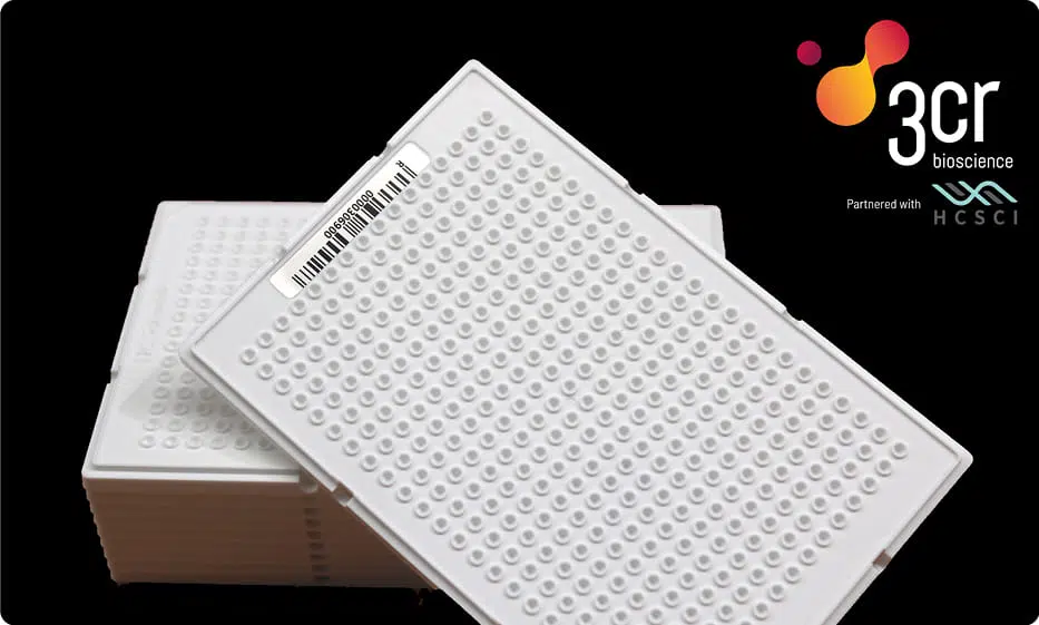
Specially designed custom low volume, low profile/slim 384-well plates designed for use with our GeneArrayer. These plates are made from durable polypropylene delivering an excellent low fluorescence background interference and high performance when paired with our high-throughput genotyping instruments.
The unique, slimline plate design permits lower reaction volumes than standard 384-well plates (typically 1.6 – 2.0 µL) allowing for reaction miniaturization, with all the benefits this brings saving time, consumables and costs. This plate is designed specifically to be used with 3CR Bio’s GeneArrayer automated liquid handling instrument.
Plate Dimensions: Length 127.85 mm, Width 85.85 mm, Thickness 5.05 mm, Well Spacing 4.50 mm, Individual Well Capacity 3 µL.
Other Products
DBCO-C3-PEG4-amine
Product Info
Document
Product Info
DBCO-C3-PEG4-amine is a PEG linker which contains DBCO and amine moieties. The DBCO group is commonly used in copper-free Click Chemistry reactions. The amine group is reactive with carboxylic acids, activated NHS esters, carbonyls (ketone, aldehyde) etc. The hydrophilic PEG spacer increases solubility in aqueous media. Reagent grade, for research purpose. Please contact us for GMP-grade inquiries.
Document
DBCO-C3-PEG4-amine is a PEG linker which contains DBCO and amine moieties. The DBCO group is commonly used in copper-free Click Chemistry reactions. The amine group is reactive with carboxylic acids, activated NHS esters, carbonyls (ketone, aldehyde) etc. The hydrophilic PEG spacer increases solubility in aqueous media. Reagent grade, for research purpose. Please contact us for GMP-grade inquiries.
[PS1000] FluoroStain™ Protein Fluorescent Staining Dye (Red, 1,000X), 1 ml
Product Info
Document
Product Info
Description
The FluoroStain™ Protein Fluorescent Staining Dye (Red, 1000×) is designed to substitute the common Coomassie Blue protein staining method, offering greater sensitivity and ease of operation. Unlike Coomassie Blue stain, the FluoroStain Protein Fluorescent Staining Dye binds to protein with high specificity, making destaining process an option rather than a requirement. With further reduction of background signals via destaining process, the FluoroStain™ is capable of achieving detection level parallel to silver staining without specialized imaging equipment, making it one of the most sensitive dyes available. In addition to its remarkable sensitivity, the FluoroStain™ Protein Fluorescent Staining Dye (Red, 1000×) brings a more reliable and safer user experience, since the stained gel can be visualized with blue-light illumination, avoiding the risk of skin/ eye damage caused by UV light. For best result, we suggest using B-BOX™ Blue Light LED epi-illuminator to visualize and analyze the gel stained with FluoroStain Protein Fluorescent Staining Dye (Red, 1000×). The FluoroStain™ Protein Fluorescent Staining Dye is compatible to the analysis of mass spectra, i.e. LC-MS/MS, MALDI-TOF, etc.
Spectral Characteristics
When it is bound with bovine serum albumin (BSA), the fluorescent emission of FluoroStain Protein Fluorescent Staining Dye can be excited by UV and blue light sources, with excitation peaks around 369 and 517 nm and emission at 605 nm. In absence of BSA, FluoroStain Protein Fluorescent Staining Dye shows ignorable fluorescence as compared with protein-bound form, therefore giving a clear background for photographic analysis.
These spectral characteristics made this fluorescent dye compatible with a wide variety of gel reading facilities, including UV/ blue light epi- and transilluminator, argon laser and mercury-arc lamp excitation gel scanners.
Storage
Protected from light -20°C for 24 months
Document
The FluoroStain™ Protein Fluorescent Staining Dye (Red, 1000×) is designed to substitute the common Coomassie Blue protein staining method, offering greater sensitivity and ease of operation. Unlike Coomassie Blue stain, the FluoroStain Protein Fluorescent Staining Dye binds to protein with high specificity, making destaining process an option rather than a requirement. With further reduction of background signals via destaining process, the FluoroStain™ is capable of achieving detection level parallel to silver staining without specialized imaging equipment, making it one of the most sensitive dyes available. In addition to its remarkable sensitivity, the FluoroStain™ Protein Fluorescent Staining Dye (Red, 1000×) brings a more reliable and safer user experience, since the stained gel can be visualized with blue-light illumination, avoiding the risk of skin/ eye damage caused by UV light. For best result, we suggest using B-BOX™ Blue Light LED epi-illuminator to visualize and analyze the gel stained with FluoroStain Protein Fluorescent Staining Dye (Red, 1000×). The FluoroStain™ Protein Fluorescent Staining Dye is compatible to the analysis of mass spectra, i.e. LC-MS/MS, MALDI-TOF, etc.
Annexin A1 (ANXA1) is a membrane protein that plays a role in innate and adaptive immunity by controlling the biosynthesis of inflammation, prostaglandins, and leukotriene mediators. This target is overexpressed in 97% of all samples from patients with with hairy cell leukemia, and is absent in other B-cell lymphomas. High ANXA1 expression is frequently associated with advanced stage esophageal and esophagogastric junction adenocarcinoma, and is also linked to advanced and metastatic disease states.