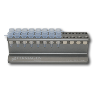
Permagen’s 0.2 mL PCR Strip Magnetic rack is designed for magnetic bead separations from Individual tubes, 8-strip, or 12-strip PCR tubes
Detail
Permagen’s 0.2 mL PCR Strip Magnetic rack is designed for magnetic bead separations from Individual tubes, 8-strip, or 12-strip PCR tubes
Accommodates any common PCR strips or individual PCR tubes
Tubes are angled and beads will be pulled to back wall allowing easy aspiration and tip tracking down the front wall of the tubes without disturbing bead pellet
Bring your assay kits to the next level with freeze-dried PCR beads.
LyoBeads are ready-to-use, freeze-dried master mixes in shape of a small ball or spheres.
LyoBeads contain: DNA polymerase(s), reaction buffer, dNTPs and primers and probes. They are rehydrated within seconds in any aqueous solutions, which makes reaction setup very simple. Only a biological sample has to be added.
Storage: LyoBeads are shipped and stored simply at room-temperature. This provides a more cost efficient and ecological distribution compared to other master mixes.
LyoBeads can be pre-dispensed in PCR-strips, PCR-plates, cartridges etc.
As this is a customized product, we take your requirements very serious. You would like to know more? Please get in touch!
Document
Bring your assay kits to the next level with freeze-dried PCR beads.
LyoBeads are ready-to-use, freeze-dried master mixes in shape of a small ball or spheres.
LyoBeads contain: DNA polymerase(s), reaction buffer, dNTPs and primers and probes. They are rehydrated within seconds in any aqueous solutions, which makes reaction setup very simple. Only a biological sample has to be added.
endo-BCN-PEG3-Gly represents an ADC linker featuring a BCN group, a hydrophilic PEG3 linker, and glycine residue. The BCN group is a click chemistry handle that readily reacts with azide groups on a target molecule to form stable triazole linkages. The PEG units enhance the aqueous solubility of the compound and may assist in fine-tuning DMPK properties. This compound may be applied to the synthesis of antibody-drug conjugates.
Document
endo-BCN-PEG3-Gly represents an ADC linker featuring a BCN group, a hydrophilic PEG3 linker, and glycine residue. The BCN group is a click chemistry handle that readily reacts with azide groups on a target molecule to form stable triazole linkages. The PEG units enhance the aqueous solubility of the compound and may assist in fine-tuning DMPK properties. This compound may be applied to the synthesis of antibody-drug conjugates.
African Swine Fever Virus (ASFV) is a widespread disease which infects members of the pig family(Suidae). Anumberoftick species are believed to be the vector for the disease,as well as being transmitted by raw pork and pig excrement [1]. After firstly being identified in Kenya in 1921, ASFV became endemic in sub-Saharan Africa, with regular outbreaks being reported across Europe, Asia and South America throughout the century [2]. More recently the virus was introduced in Georgia and spread throughout the region, as well as mass outbreaks occurring in China in 2018 [3]. ASFVistheonlymemberoftheAsfaridaefamily.ItisalargeenvelopeddoublestrandedDNA virus of icosahedral morphology with an average diameter of 200nm and isolates contain genomes between 170-190Kbp encoding for up to 167 open reading frames [2]. The morphology of ASFV consist of several concentric domains. An inner core contains the nucleoid coated with a thick protein layered core shell, which is surrounded by an inner lipid envelope , all of which is encompassed by the capsid [2]. ASFV begins its replication cycle in the nucleus of infected cells before moving to the cytoplasm where the majority of the replication takes place [2]. Gene transcription is highly regulated, with distinct classes of mRNA identified to accumulate at early, intermediate and late transcripts of the virus [2]. The disease induces acute haemorrhagic disease within its hosts, causing high fevers and skin haemorrhages, with death often occurring within ten days of clinical symptoms appearing [4].
References: 1: The Centre for Food Security and Public Health (2015), African Swine Fever. 2: Galindo, I. and Alonso, C., 2017. African swine fever virus: a review. Viruses, 9(5), p.103. 3: Zhou, X., Li, N., Luo, Y., Liu, Y., Miao, F., Chen, T., Zhang, S., Cao, P., Li, X., Tian, K. and Qiu, H.J., 2018. Emergence of African swine fever in China, 2018. Transboundary and emerging diseases, 65(6), pp.1482-1484. 4: Gallardo, C., Ademun, A.R., Nieto, R., Nantima, N., Arias, M., Martín, E., Pelayo, V. and Bishop, R.P., 2011. Genotyping of African swine fever virus (ASFV) isolates associated with disease outbreaks in Uganda in 2007. African Journal of biotechnology, 10(17), pp.3488-3497.
Document
Exceptional value for money Rapid detection of all clinically relevant subtypes Positive copy number standard curve for quantification Highly specific detection profile High priming efficiency Broad dynamic detection range (>6 logs) Sensitive to < 100 copies of target
Accurate controls to confirm findings