RPA/MIRA is used for DNA and RNA nucleic acid templates isothermal amplification , and can be used in the field of molecular detection of viruses, pathogenic bacteria, tissues, cells, etc.
Detail
Payment & Shipping Terms
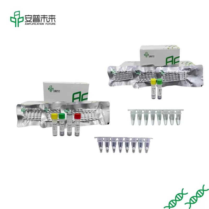
Packaging Details
16T/Bag,48T/Box
Delivery Time
6 working days
Payment Terms
T/T, MoneyGram
Supply Ability
100000T/Month
Product Description
RPA/MIRA is used for DNA and RNA nucleic acid templates isothermal amplification , and can be used in the field of molecular detection of viruses, pathogenic bacteria, tissues, cells, etc.
MIRA VS PCR ,MIRA Advantages:
Low and constant temperature, low requirements on instruments
20mins
Only one pair of primers is needed
Fluorescence detection by probe method is also available
High specificity
PCR :Need to control temperature,90 minutes
LAMP:Design three pairs of primers,60minutes,Low specificity
Other Products
NGS DNA Library Prep Kit (Ion Torrent Platform)
Product Info
Document
Product Info
The NGS DNA Library Prep Kit (Ion Torrent platform) was developed for construction of high quality DNA libraries for next generation sequencing using Ion Torrent platform. The kit uses short double strand DNA fragments (blunt ends and/or sticky ends) as input DNA for NGS library construction, and is compatible with DNA fragments generated from both enzymatic methods (BioDynami DNA Fragmentation Enzyme Mix and DNA Fragmentation & A-tailing Enzyme Mix etc) and mechanical methods (sonication, nebulization etc.). Our unique technology increases library conversion efficiency and eliminates insert concatemer ligation. Library multiplexing up to 12 samples is possible.
NGS DNA Library Prep Kit Workflow
Kit features
Simple procedure: Reactions for all three steps are in one tube
Fast protocol
Total protocol time is around 1 hour
Hands-on time is only ~5 minutes
Guaranteed high library conversion efficiency as compared to other kits
Input DNA amount: from 50 ng to 1 ug
No insert concatemer ligation
Document
The NGS DNA Library Prep Kit (Ion Torrent platform) was developed for construction of high quality DNA libraries for next generation sequencing using Ion Torrent platform. The kit uses short double strand DNA fragments (blunt ends and/or sticky ends) as input DNA for NGS library construction, and is compatible with DNA fragments generated from both enzymatic methods (BioDynami DNA Fragmentation Enzyme Mix and DNA Fragmentation & A-tailing Enzyme Mix etc) and mechanical methods (sonication, nebulization etc.). Our unique technology increases library conversion efficiency and eliminates insert concatemer ligation. Library multiplexing up to 12 samples is possible.
Plasmid Purification Magnetic Beads (RNA Depletion)
Product Info
Document
Product Info
Plasmid Purification Magnetic Beads (RNA Depletion)
Plasmid isolation from bacterial cultures is one of the most popular techniques in biomedical research and pharmaceutical industries. However, it is common that the isolated plasmid DNA is usually contaminated with varied degrees of host RNA. Plasmid purification is necessary to reduce the impact on downstream applications by removing RNA contamination.
We have developed a simple reagent to completely remove RNA contamination in the isolated plasmid samples using Solid Phase Reversible Immobilization (SPRI) beads. SPRI beads consist of paramagnetic particles coated with carboxyl groups that reversibly bind DNA. Our Plasmid Purification Magnetic Beads (RNA Depletion) combines BioDynami’s proprietary chemistries with the reversible DNA-binding properties of SPRI magnetic beads. The reagent removes RNA and recovers the plasmid in the same step. Moreover, unwanted components such as salts, dNTPs, proteins, enzymes, and other impurities can also be removed simultaneously.
Plasmid can be used for downstream applications such as enzymatic digestion, transformation, transfection and molecular cloning etc. The beads can be an effective and inexpensive reagent for bacterial RNA depletion for routine plasmid purification.
Features
Effective depletion of bacterial RNA by RNase
High recovery rate of plasmid DNA by magnetic beads
Removal of unwanted components and impurities
Simple and fast beads-based protocol
Document
Plasmid isolation from bacterial cultures is one of the most popular techniques in biomedical research and pharmaceutical industries. However, it is common that the isolated plasmid DNA is usually contaminated with varied degrees of host RNA. Plasmid purification is necessary to reduce the impact on downstream applications by removing RNA contamination.
[PM2500] ExcelBand™ 3-color Regular Range Protein Marker (9-180 kDa), 250 μl x 2
Product Info
Document
Product Info
Description
The PM2500 ExcelBand™ 3-color Regular Range Protein Marker is a ready-to-use three-color protein standard with 10 pre-stained proteins covering a wide range of molecular weights from 10 to 180 kDa in Tris-Glycine Buffer (9 to 170 kDa in Bis-Tris (MOPS) buffer and 10 to 170 kDa Bis-Tris (MES) buffer). Proteins are covalently coupled with a blue chromophore except for two reference bands (one green and one red band at 25 kDa and 75 kDa respectively) when separated on SDS-PAGE (Tris-Glycine buffer). PM2500 ExcelBand™ 3-color Regular Range Protein Marker is designed for monitoring protein separation during SDS-polyacrylamide gel electrophoresis, verification of Western transfer efficiency on membranes (PVDF, nylon, or nitrocellulose) and for approximating the size of proteins.
Features
Ready-to-use — Premixed with a loading buffer for direct loading, no need to boil.
Two reference bands — 75 kDa (red) and 25 kDa (green)
Contents
Approximately 0.1~0.4 mg/ml of each protein in the buffer (20 mM Tris-phosphate (pH 7.5 at 25°C), 2% SDS, 0.2 mM DTT, 3.6 M urea, and 15% (v/v) glycerol).
Quality Control
Under suggested conditions, PM2500 ExcelBand™ 3-color Regular Range Protein Marker resolves 10 major bands in 15% SDS-PAGE (Tris-Glycine buffer, MOPS, and MES buffer) and after Western blotting to nitrocellulose membrane.
Storage
4°C for 3 months -20°C for long term storage
Document
The PM2500 ExcelBand™ 3-color Regular Range Protein Marker is a ready-to-use three-color protein standard with 10 pre-stained proteins covering a wide range of molecular weights from 10 to 180 kDa in Tris-Glycine Buffer (9 to 170 kDa in Bis-Tris (MOPS) buffer and 10 to 170 kDa Bis-Tris (MES) buffer). Proteins are covalently coupled with a blue chromophore except for two reference bands (one green and one red band at 25 kDa and 75 kDa respectively) when separated on SDS-PAGE (Tris-Glycine buffer). PM2500 ExcelBand™ 3-color Regular Range Protein Marker is designed for monitoring protein separation during SDS-polyacrylamide gel electrophoresis, verification of Western transfer efficiency on membranes (PVDF, nylon, or nitrocellulose) and for approximating the size of proteins.