
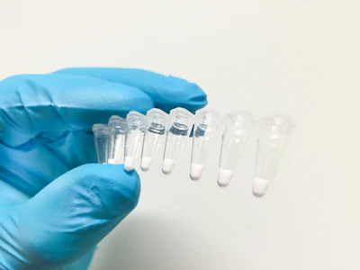

LyoCakes are ready-to-use, freeze-dried master mixes pre-aliquoted PCR-strips or reaction-tubes.
LyoCakes contain: DNA polymerase(s), reaction buffer, dNTPs and primers and probes. They are rehydrated within seconds in any aqueous solutions, which makes reaction setup very easy. Simply add a biological sample and place the tube in a PCR cycler.
Storage: LyoCakes are shipped and stored simply at room-temperature. This provides a more cost efficient and ecological distribution compared to other master mixes.
As this is a customized product, we take your requirements very serious. You would like to know more? Please get in touch!


