
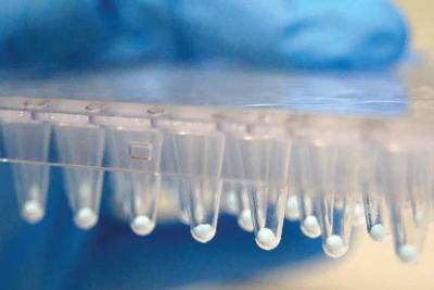

LyoBeads are ready-to-use, freeze-dried master mixes in shape of a small ball or spheres.
Standard LyoBeads contain DNA polymerase(s), reaction buffer and dNTPs. LyoBeads can also contain Primer & Probes when produced customized. They are rehydrated within seconds in any aqueous solutions, which makes reaction setup very simple. Only a biological sample has to be added.
Storage: LyoBeads are shipped and stored simply at room-temperature. This provides a more cost efficient and ecological distribution compared to other master mixes.
LyoBeads can be pre-dispensed in PCR-strips, PCR-plates, cartridges etc.
All necessary components for PCR are already included in one LyoBead: an engineered DNA polymerase, an optimized reaction buffer and ultrapure dNTPs. Only primers and probes need to be added. A hot-start formulation of the included DNA polymerase prevents false amplification during the reaction set-up.
For research use and further manufacturing.
In case you are aiming to use our RUO products as components or for your development of e.g. an IVD medical device, please contact us.


