PRP Centrifuge widly used for Aesthetic & Plastic,Orthopedic & Pain-treatment, Dentistry, Ophthalmology,Veterinary and etc..Our company has been cooperated with Korean and Chinese PRP kit manufacturer to making the PRP centrifuge to our customers
Detail
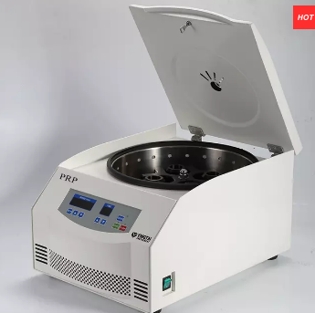
PRP centrifuge/Platelet rich plasma centrifuge
PRP Application:
Aesthetic & Plastic,Orthopedic & Pain-treatment, Dentistry, Ophthalmology,Veterinary and etc..Our company has been cooperated with Korean and Chinese PRP kit manufacturer to making the PRP centrifuge to our customers.Detail information, please send email to us, [email protected]
Features:
1.Widely used in hospital, modern beauty salon and micro plastic hospitals.
DC inverter motor with simpler construction, more reliable performance, longer life and quietly running
2.Smooth in operation, low noise and small vibration.
3.Micro computer control system, digital display the RCF, time and speed. 4.Automatic electric lid, compact design, super speed and imbalance protection.
5.The centrifuge body is made of high-quality steel, safe and reliable.
TD4N Technical Parameters:
Max speed
4000rpm
Max RCF
2600xg
Max volume
4x20ml
Noise
≤55dBA
Timer
0~99min
Net weight
28KG
Dimension(HxDxW)
528×370×280mm
Power supply
AC 220V 50HZ 2A
Speed accuracy
±20rpm
Package
Carton box
Other Products
Cell Culture Flask T175
Product Info
Document
Product Info
Cell Culture Flask T175
Cell Culture Flask surface is very smooth based on precise molding technology and it gives clear view when examined with microscope.
Volume: 25mL75mL175mL225mL
PRODUCT FEATURES
The product is made of medical grade USP CLASS VI polymer polystyrene
The product is made under a 100,00- class dust-free manufacturing site
Two kinds of product line up are providing.
For adherent cell culture: Initial adherence and proliferative property of cells via hydrophilic surface treatment.
For suspension cell culture: The surface is resistant to cell adherence, which minimizes damage or loss of cell.
Large mouthed design makes easy operation of pipet or cell scraper. The surface of flask is uniform and smooth, hence the clear view can be obtained when microscopic observation.
The hydrophobic filter cap can prevent invasion of fungi and bacteria without water absorb.
Gamma radiation sterilization
Non Byrogenic Nase/Bnase free
Document
Cell Culture Flask surface is very smooth based on precise molding technology and it gives clear view when examined with microscope.
For the selective isolation and culture of Listeria monocytogenes.
Principle and Interpretation
Peptone provides the carbon and nitrogen sources necessary for growth; yeast extract powder and starch provide carbon and nitrogen sources, vitamins and growth factors; sodium chloride can maintain a balanced osmotic pressure; glucose provides a carbon source; Listeria hydrolyzes esculin and reacts with iron ions to form black 6,7-dihydroxycoumarin; mannitol is a fermentable sugar, and phenol red is a pH indicator; lithium chloride and other antibiotics can inhibit Gram-negative bacteria and most Gram-positive bacteria; agar is the coagulant of the culture medium.
Formulation
Ingredients
/liter
Peptone
23 g
Starch
1 g
NaCl
5 g
Columbia agar
13 g
Mannitol
10 g
Ferric ammonium citrate
0.5 g
Esculin (aesculin)
0.8 g
Dextrose (glucose)
0.5 g
Lithium chloride
15.0 g
Phenol red
0.08 g
pH7.2±0.2 at 25°C
Preparation
Weigh 68.9g of dry powder of this product, add 1L of distilled water or deionized water, stir, heat and boil until completely dissolved, divide into Erlenmeyer bottles, sterilize at 121℃ for 15min, cool to room temperature and set aside. Heat to dissolve agar before use, cool to 50℃, add 1 tube of supporting reagent (SR0140) per 100mL of basal culture medium, shake well and pour into sterilized culture dish.
Quality Control
The following quality control strains were inoculated and cultured at 35-37℃ for 24h. The results are as follows:
Quality control strains
Growth
Listeria monocytogenes ATCC19115
Gray-green colonies with a black depression in the center and black surrounding
Enterococcus faecalis ATCC29212
–
Escherichia coli ATCC25922
–
Storage and Shelf Life
2-30℃,Keep container tightly closed, avoid direct sunlight.
Use before expiry date on the label.
Precautions
1. When weighing the dehydrated medium, please wear masks to avoid causing respiratory system discomfort
2. Keep container tightly closed after using to prevent clumping.
Waste Disposal
Microbiological contamination was disposed by autoclaving at 121°C for 30 minutes.
Document
Intended Use For the selective isolation and culture of Listeria monocytogenes. Principle and Interpretation Peptone provides the carbon and nitrogen sources necessary for growth; yeast ex……
Rapid isolation of both small and large species of DNA from urine
Convenient spin column format
Effective removal of PCR inhibitors
Purified DNA is highly suited to sensitive downstream applications
Allows for the purification of viral DNA from urine
Both high molecular weight DNA (greater than 1 kb in size; mostly cell associated) and the smaller DNA (150 – 250 bp; derived from the circulation) is effectively isolated and purified using a rapid and convenient spin column protocol. This kit can be used to isolate DNA from a broad range of viruses in urine as well. Salts, metabolic wastes, proteins and other contaminants are removed to yield inhibitor-free DNA for use in sensitive applications. The DNA is of excellent quality for various downstream applications such as PCR, qPCR and DNA fingerprinting, methylation studies and more.
This kit provides a fast, reliable and simple procedure for isolating DNA from urine volumes ranging from 50 μL to 1.75 mL of urine. Multiple samples can be processed in 30 minutes.
Urine DNA Isolation Kit (Slurry Format)
This kit provides a fast, reliable and simple procedure for isolating DNA from urine volumes ranging from 3 mL to 25 mL. Multiple samples can be processed in 30 minutes. Multiple samples can be processed in 45 minutes.
Urine DNA Isolation Maxi Kit (Slurry Format)
This kit provides a fast, reliable and simple procedure for isolating DNA from urine volumes ranging from 25 mL of urine up to 80 mL. Multiple samples can be processed in 45 minutes.
Background
DNA found in urine can be divided into 2 basic categories. The larger species, genomic-DNA (gDNA), is generally greater than 1 kb in size, and appears to be derived mainly from exfoliated cells. The second species is smaller, generally between 150 and 250 bp (apoptotic-DNA), and derives, at least in part, from the circulation. The second species is also considered as an RNA/DNA hybrid as reported by Halicka et al. (2000). Both types of DNA can be isolated reliably using this kit.
Storage Conditions and Product Stability All buffers should be kept tightly sealed and stored at room temperature. This kit is stable for 2 years after the date of shipment. It is recommended to warm up Lysis Buffer A for 20 minutes at 60°C if any salt precipitation is observed.