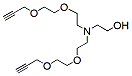
Hydroxy-Amino-bis(PEG2-propargyl) is a 3-arm linker with a terminal hydroxy group and two propargyl groups. The propargyl groups can participate in copper catalyzed azide-alkyne Click Chemistry to yield a stable triazole linkage. Reagent grade, for research purpose. Please contact us for GMP-grade inquiries.
Detail
Hydroxy-Amino-bis(PEG2-propargyl) is a 3-arm linker with a terminal hydroxy group and two propargyl groups. The propargyl groups can participate in copper catalyzed azide-alkyne Click Chemistry to yield a stable triazole linkage. Reagent grade, for research purpose. Please contact us for GMP-grade inquiries.
Other Products
N-(t-butyl ester-PEG2)-N-bis(PEG2-propargyl)
Product Info
Document
Product Info
N-(t-butyl ester-PEG2)-N-bis(PEG2-propargyl) is a multi-functional PEG linker with two terminal propargyl groups and a t-butyl ester. The propargyl groups enables formation of triazole linkage with azide-bearing compounds or biomolecules in copper catalyzed Click Chemistry. The t-butyl protected carboxyl group can be deprotected under mild acidic conditions. Reagent grade, for research purpose. Please contact us for GMP-grade inquiries.
Document
N-(t-butyl ester-PEG2)-N-bis(PEG2-propargyl) is a multi-functional PEG linker with two terminal propargyl groups and a t-butyl ester. The propargyl groups enables formation of triazole linkage with azide-bearing compounds or biomolecules in copper catalyzed Click Chemistry. The t-butyl protected carboxyl group can be deprotected under mild acidic conditions. Reagent grade, for research purpose. Please contact us for GMP-grade inquiries.
This product supplies a simple and rapid extraction of total RNA from tissue and culture cells samples. The kit is based on superparamagnetic particles purification technology, no phenol-chloroform extraction or alcohol precipitation. The whole extraction processis only 60 minutes. Purified RNA is ready for downstream applications such as RT-PCR, virus RNA testing and so on. MagPure LQ Kit buffers can be used for both manual extraction process and automatic nucleic acid extraction machines.
Details
Specifications
Features
Specifications
Main Functions
Isolation total RNA from tissue and cells and plant
Applications
RT-PCR, cDNA synthesis, second generation sequencing
Purification method
Polydisperse magnetic beads
Purification technology
Magnetic beads technology
Process method
Manual or automatic
Adaptive instrument
Nucleic acid extractor, pipetting workstation
Sample type
Animal tissue, human tissue, cultured cells, lymphocytes, ordinary plant tissue
Sample amount
Cells: 1 x 10⁷Animal tissue: ≤20mgPlant tissue: ≤100mgYeast cell: ≤5 x 10⁶
Yield
5-100μg
Principle
The Kit combines the speed and efficiency of silica-based technology with the convenient handling of magnetic particles for purification of total RNA. Samples are lysed and RNA is purified from lysates in one step through its binding to the silica surface of the particles in the presence of a chaotropic salt. The particles are separated from the lysates using a magnet and DNA is removed by treatment with RNase-free DNase. The magnetic particles are efficiently washed, and RNA is eluted in RNase-free water.
Advantages
High purity – OD 260 / 280 :1.9-2.0, OD 260 / 230 :1.5-2.0
Economy – less than 50% of the price of Qiagen and other imported products
Automatic – extraction without manual participation, saving time and effort
Universal – suitable for various animal tissues, cultured cells and conventional plant fungal tissues
Kit Contents
Contents
R662201
R662202
R662203
Purification Times
48 Preps
96 Preps
5 x 96 Preps
MagPure RNA Particles
1.7 ml
4.0 ml
18 ml
Proteinase K
24 mg
50 mg
240 mg
Protease Dissolve Buffer
1.8 ml
5 ml
15 ml
DNase I
600 μl
2 x 600 μl
10 x 600 μl
DNase Buffer
30 ml
40 ml
200 ml
RTL Lysis Buffer
40 ml
80 ml
400 ml
Buffer MCB*
18 ml
30 ml
150 ml
Buffer MW1*
44 ml
66 ml
2 x 220 ml
Buffer RW2*
20 ml
50 ml
2 x 100 ml
RNase Free Water
10 ml
30 ml
120 ml
Storage and Stability
MagPure RNA Particles and Proteinase K should be stored at 2–8°C upon arrival. DNase I shouldbe stored at -20°C. However, short-term storage (DNase I up to 1 weeks, MagPure RNA Particles and Proteinase K up to 8 weeks) at room temperature (15–25°C) does not affect their performance. The remaining kit components can be stored at room temperature (15–25°C) and are stable for atleast 18 months under these conditions.
Document
This product supplies a simple and rapid extraction of total RNA from tissue and culture cells samples. The kit is based on superparamagnetic particles purification technology, no phenol-chloroform extraction or alcohol precipitation. The whole extraction processis only 60 minutes. Purified RNA is ready for downstream applications such as RT-PCR, virus RNA testing and so on. MagPure LQ Kit buffers can be used for both manual extraction process and automatic nucleic acid extraction machines.
Propargyl-PEG12-methane is a PEG linker that can react with azide-bearing compounds or biomolecules via copper catalyzed Click Chemistry to form a stable triazole linkage. The PEG chain enhances the solubility of the molecule in aqueous media. Reagent grade, for research purpose. Please contact us for GMP-grade inquiries.
Document
Propargyl-PEG12-methane is a PEG linker that can react with azide-bearing compounds or biomolecules via copper catalyzed Click Chemistry to form a stable triazole linkage. The PEG chain enhances the solubility of the molecule in aqueous media. Reagent grade, for research purpose. Please contact us for GMP-grade inquiries.