
Description
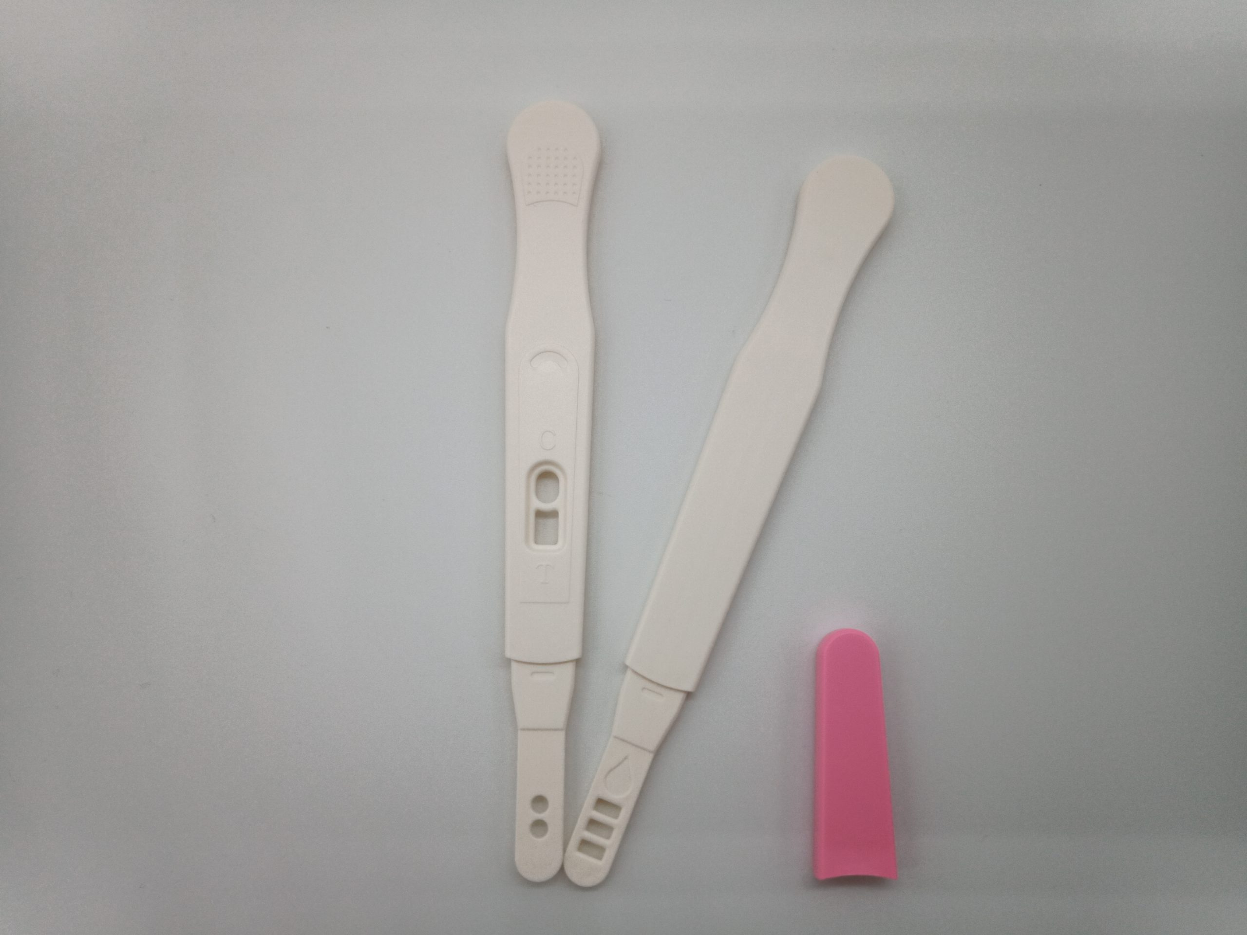
- Appearance: White/pink
- • Length of strip: 86mm
- • Width of strip: 4.5mm
- • Length of device: 136mm
- • Width of device: 16mm


Norgen’s EXTRAClean Urine Exosome Purification and RNA Isolation Mini Kit constitutes an all-in-one system for the purification of exosomes and the subsequent isolation of RNA from different urine sample volumes ranging from 250 μL to 1 mL. The purification is based on spin column chromatography that employs Norgen’s proprietary resin. The EXTRAClean columns undergo stringent processing and rigorous quality control measures to minimize contamination traces, ensuring optimal results for sensitive applications such as NGS. The kit is designed to isolate all sizes of RNA, including microRNA. The kit provides a clear advantage over other available kits in that it does not require any special instrumentation, protein precipitation reagents, extension tubes, phenol/chloroform or protease treatments. Moreover, the kit allows the user to elute into a flexible elution volume ranging from 50 μL to 100 μL. The purified RNA is free from any protein-bound circulating RNA and of the highest integrity. The purified RNA can be used in a number of downstream applications including real time PCR, reverse transcription PCR, NGS application, Northern blotting, RNase protection and primer extension, and expression array assays.
Figure 1 / 9

Click for expanded view
| Minimum Urine Input | 2 mL |
| Maximum Urine Input | 10 mL |
| Size of Exosomes Purified | 40 nm – 150 nm |
| Size of RNA Purified | All sizes, including miRNA and small RNA (< 200 nt) |
| Elution Volume | 50-100 μL |
| Time to Complete 10 Purifications | 35-40 minutes |
| Average Yields | Variable depending on specimen |
Storage Conditions and Product Stability
All buffers should be kept tightly sealed and stored at room temperature. This kit is stable for 2 years after the date of shipment. It is recommended to warm Lysis Buffer A for 20 minutes at 60°C if any salt precipitation is observed.

Description
The ExcelTaq™ 2X Q-PCR Master Mix (SYBR, ROX) is a ready-to-use reagent with all the essential components for quantitative real-time PCR (qPCR) except primers and template. The master mix features high sensitivity and high signal intensity when compared with Brand A. The ExcelTaq™ 2X Q-PCR Master Mix (SYBR, ROX) contains hot-start Taq polymerase in an optimized buffer with dsDNA specific SYBR green fluorescent dye. This master mix allows for sensitive and precise amplification, real-time tracking of the amplification process, and simultaneous quantification for targeted DNA molecules. The master mix includes ROX reference dye for normalization of each qPCR assay. With inert smart blue contrast dye, the ExcelTaq™ 2X Q-PCR Master Mix (SYBR, ROX) is ready-to-use and greatly reduces pipetting errors, while largely improving the reproducibility of the process.
Features
Storage
Protected from light.
Aliquot to avoid multiple freeze-thaw cycles.
-20°C for 12 months
The ExcelTaq™ 2X Q-PCR Master Mix (SYBR, ROX) is a ready-to-use reagent with all the essential components for quantitative real-time PCR (qPCR) except primers and template. The master mix features high sensitivity and high signal intensity when compared with Brand A. The ExcelTaq™ 2X Q-PCR Master Mix (SYBR, ROX) contains hot-start Taq polymerase in an optimized buffer with dsDNA specific SYBR green fluorescent dye. This master mix allows for sensitive and precise amplification, real-time tracking of the amplification process, and simultaneous quantification for targeted DNA molecules. The master mix includes ROX reference dye for normalization of each qPCR assay. With inert smart blue contrast dye, the ExcelTaq™ 2X Q-PCR Master Mix (SYBR, ROX) is ready-to-use and greatly reduces pipetting errors, while largely improving the reproducibility of the process.

Nucleic acid testing (NAT) is the method of choice for detection and quantification of a wide range of micro organisms. Primerdesign manufactures and supplies high quality quantitative real-time PCR kits for the detection and simultaneous quantification of numerous significant pathogens . A copy number standard curve is provided for quantification and an the internal extraction template (DNA or RNA), controls for the quality of the nucleic acid extraction and eliminates false negative results.
The kit is designed with the broadest possible detection profile to ensure that all clinically relevant strains and subtypes are detected. Target sequences are selected by working with data from key opinion leaders in the field. Multiple sequence alignments and unprecedented real-time PCR expertise in design and validation ensure the best possible kit. Details of the target and priming specificity are included in the individual handbooks above.
Packaged, optimised and ready to use. Expect Better Data.
Exceptional value for money
Rapid detection of all clinically relevant subtypes
Positive copy number standard curve for quantification
Highly specific detection profile
High priming efficiency
Broad dynamic detection range (>6 logs)
Sensitive to < 100 copies of target
Accurate controls to confirm findings
150 reactions