Usages: 
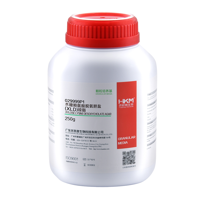
For selective isolation of Gram-negative bacteria, especially for Shigella and Salmonella.
Principle: Yeast extract powder provide nitrogen, vitamins, growth factors; sodium chloride to maintain osmotic equilibrium ferric ammonium citrate of iron salts to produce a black iron sulfide; agar as medium coagulant; phenol red as pH indicator.
Formulation(per liter): Xylose 3.50g L-Lysine 5.00g Lactose 7.50g Sucrose 7.50 g Sodium Chloride 5.00 g Yeast Extract 3.00g Sodium Desoxycholate 2.50g Sodium Thiosulphate 6.80g Fe-Ammonium Citrate 0.80g Phenol Red 0.08g Agar 13.50 Final PH 7.4±0.2
How to use: 1.Suspend 55.2g in 1Lof distilled or deionized water. Heat with frequent agitation and boil to completely dissolve the powder. Do not autoclave ,cool to 50℃ and pour into sterile petri dishes. 2.Dilluted and treated samples.
Quality control: Quality control strains were inoculated ,and cultured at 36 ± 1 ℃ for 24h ,results show as follows: strain name strain code growth feature Arizona bacteria CMCC (B) 47001 good black colonies Salmonella typhimurium CMCC (B) 50115 good black colonies Streptococcus faecalis CMCC32223 prohibited —
Storage: Store in a dark, cool and dry place, tighten the cap immediately after use. Storage period of three years.
Specifications: 250g/bottle
Other Products
Clostridium difficile TaqMan PCR Lyophilized Kits
Product Info
Document
Product Info
Overview
Detection kits for Clostridium difficile
Lyophilized format designed for room temperature shipping
Available in TaqMan format for analysis
Norgen’s Clostridium difficile TaqMan PCR Lyophilized Kit is designed for the detection of Clostridium difficile specific DNA in a real-time PCR based on the use of TaqMan® technology. The lyophilized format is designed to ship the kit at ambient temperature.
Norgen’s Clostridium difficile TaqMan Lyophilized Probe/Primer and Control Set is designed for the detection of Clostridium difficile specific DNA in a real-time PCR based on the use of TaqMan® technology. The lyophilized format is designed to ship the kit at ambient temperature.
All kit components should be stored at -20°C upon arrival.
Once reconstituted, repeated thawing and freezing (>2 times) of the Master Mix and Positive Control should be avoided, as this may affect the performance of the assay. If the reagents are to be used only intermittently, they should be frozen in aliquots.
Each kit is provided with 4 tubes of 2X PCR Master Mix and each tube is enough to run 25 reactions. It is not necessary to reconstitute all Master Mix tubes at once. The Master Mix tubes can be reconstituted as and when needed.
All kit components can be stored for 2 years after the date of production without showing any reduction in performance.
HiPure Insect DNA Kits provides a simple and rapid solution for total DNA extraction of insect tissue samples. This kit is based on silica gel column purification technology without toxic phenol chloroform extraction and time-consuming alcohol precipitation. The whole extraction process only takes 30 minutes. HiPure Insect DNA Kit can process tissue samples less than 10mg at a time. Hipure Insect DNA 96 kit can process 96 insect tissue samples at a high throughput. The obtained DNA can be directly used in PCR, Southern blot, viral DNA detection and other experiments.
Details
Specifications
Features
Specifications
Main Functions
Isolation total DNA from insect tissue
Applications
PCR, southern bolt and virus detection, etc
Purification method
96 well DNA plate
Purification technology
Silica technology
Process method
Manual (centrifugation or vacuum)
Sample type
Insect tissue samples
Sample amount
Elution volume
Time per run
Liquid carrying volume per column
Binding yield of column
Principles
This product is based on silica column purification. The sample is lysed and digested with lysate and protease, DNA is released into the lysate. Transfer to an adsorption column. Nucleic acid is adsorbed on the membrane, while protein is not adsorbed and is removed with filtration. After washing proteins and other impurities, Nucleic acid was finally eluted with low-salt buffer (10mm Tris, pH9.0, 0.5mm EDTA).
Advantages
High purity DNA – can be used in sensitive downstream applications such as multiplex and quantitative pcr
Good repeatability – suitable for extracting high-yield DNA from insect tissue samples
Safe – without phenol chloroform extraction and alcohol precipitation
Kit Contents
Contents
D313901
D313902
Purification Times
1 x 96
4 x 96
HiPure DNA Plate
1
4
2.2 ml Collection Plate
1
4
1.6 ml Collection Plate
1
4
0.5ml Collection Plate
1
4
Seal Film
8
32
Buffer ITL
30 ml
120 ml
Buffer IL
30 ml
125 ml
Buffer GW1
44 ml
2 x 110 ml
Buffer GW2
50 ml
3 x 50 ml
Proteinase K
50 mg
200 mg
Protease Dissolve Buffer
6 ml
15 ml
Buffer AE
20 ml
60 ml
Storage and Stability
Proteinase K, RNase A should be stored at 2-8°C upon arrival. However, short-term storage (up to 12 weeks) at room temperature (15-25°C) does not affect their performance. The remaining kit components can be stored at room temperature (15-25°C) and are stable for at least 18 months under these conditions. The entire kit can be stored at 2–8°C, but in thiscase buffers should be redissolved before use. Make sure that all buffers areat room temperature when used.
Document
HiPure Insect DNA Kits provides a simple and rapid solution for total DNA extraction of insect tissue samples. This kit is based on silica gel column purification technology without toxic phenol chloroform extraction and time-consuming alcohol precipitation. The whole extraction process only takes 30 minutes. HiPure Insect DNA Kit can process tissue samples less than 10mg at a time. Hipure Insect DNA 96 kit can process 96 insect tissue samples at a high throughput. The obtained DNA can be directly used in PCR, Southern blot, viral DNA detection and other experiments.
Mucin 1 (MUC1) is a membrane-bound glycoprotein involved in a number of protective and cell-signaling functions, including cell-cell adhesion, proliferation, motility, invasion, and survival. Overexpression of MUC1 is clinically indicated in breast carcinomas, papillary thyroid carcinomas, and thymic carcinomas, and reports have named MUC1 as a useful marker for differentiating thymic carcinoma from type B3 thymoma. The expression of MUC1 is correlated with the grade of malignancy in thymic epithelial tumors, and loss of MUC1 expression has been associated with reactive gastropathy. MUC1 is not expressed in normal human epidermis, but has been detected in the epidermis of psoriatic plaques of biopsies from patients diagnosed with psoriasis vulgaris.