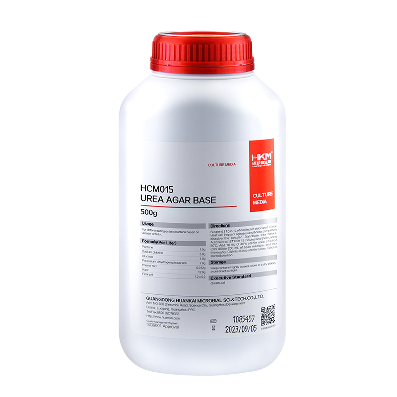
Usages: For differentiating enteric bacteria based on urease activity by adding 40% sterile urea solution.
Principle: Peptone provides the carbon and nitrogen; maintain a balanced osmotic sodium chloride; potassium dihydrogen phosphate is buffers; decomposing bacteria urease urea medium, produce large amounts of ammonia, agar as medium coagulant.
Formulation (per liter): Peptone 1g Sodium chloride 5g Glucose 1g Ppotassium dihydrogen phosphate 2g Phenol red 0.012g Agar 12g Final pH7.2 ± 0.2
How to use: 1.Suspend 21g in 1L of distilled or deionized water. Heat with frequent agitation and boil to completely dissolve the powder. Distribute into flasks. Autoclave at 121 for 15 minutes. cooling to 50-55 and adding 40% urea solution. 2.Diluted and treated samples.
Storage: Store in a dark, cool and dry place, tighten the cap immediately after use. Storage period of three years.
Other Products
Propargyl-PEG3-CH2CO2H
Product Info
Document
Product Info
Propargyl-PEG3-CH2CO2H is an PEG linker with an alkyne and carboxylic acid set of functional groups. The alkyne group can participate in copper catalyzed azide-alkyne Click Chemistry to form a stable triazole linkage. The carboxylic acid can react with primary amines to form stable amide bonds; activator (e.g. EDC, or HATU) is needed.
Document
Propargyl-PEG3-CH2CO2H is an PEG linker with an alkyne and carboxylic acid set of functional groups. The alkyne group can participate in copper catalyzed azide-alkyne Click Chemistry to form a stable triazole linkage. The carboxylic acid can react with primary amines to form stable amide bonds; activator (e.g. EDC, or HATU) is needed.
[PS1000] FluoroStain™ Protein Fluorescent Staining Dye (Red, 1,000X), 1 ml
Product Info
Document
Product Info
Description
The FluoroStain™ Protein Fluorescent Staining Dye (Red, 1000×) is designed to substitute the common Coomassie Blue protein staining method, offering greater sensitivity and ease of operation. Unlike Coomassie Blue stain, the FluoroStain Protein Fluorescent Staining Dye binds to protein with high specificity, making destaining process an option rather than a requirement. With further reduction of background signals via destaining process, the FluoroStain™ is capable of achieving detection level parallel to silver staining without specialized imaging equipment, making it one of the most sensitive dyes available. In addition to its remarkable sensitivity, the FluoroStain™ Protein Fluorescent Staining Dye (Red, 1000×) brings a more reliable and safer user experience, since the stained gel can be visualized with blue-light illumination, avoiding the risk of skin/ eye damage caused by UV light. For best result, we suggest using B-BOX™ Blue Light LED epi-illuminator to visualize and analyze the gel stained with FluoroStain Protein Fluorescent Staining Dye (Red, 1000×). The FluoroStain™ Protein Fluorescent Staining Dye is compatible to the analysis of mass spectra, i.e. LC-MS/MS, MALDI-TOF, etc.
Spectral Characteristics
When it is bound with bovine serum albumin (BSA), the fluorescent emission of FluoroStain Protein Fluorescent Staining Dye can be excited by UV and blue light sources, with excitation peaks around 369 and 517 nm and emission at 605 nm. In absence of BSA, FluoroStain Protein Fluorescent Staining Dye shows ignorable fluorescence as compared with protein-bound form, therefore giving a clear background for photographic analysis.
These spectral characteristics made this fluorescent dye compatible with a wide variety of gel reading facilities, including UV/ blue light epi- and transilluminator, argon laser and mercury-arc lamp excitation gel scanners.
Storage
Protected from light -20°C for 24 months
Document
The FluoroStain™ Protein Fluorescent Staining Dye (Red, 1000×) is designed to substitute the common Coomassie Blue protein staining method, offering greater sensitivity and ease of operation. Unlike Coomassie Blue stain, the FluoroStain Protein Fluorescent Staining Dye binds to protein with high specificity, making destaining process an option rather than a requirement. With further reduction of background signals via destaining process, the FluoroStain™ is capable of achieving detection level parallel to silver staining without specialized imaging equipment, making it one of the most sensitive dyes available. In addition to its remarkable sensitivity, the FluoroStain™ Protein Fluorescent Staining Dye (Red, 1000×) brings a more reliable and safer user experience, since the stained gel can be visualized with blue-light illumination, avoiding the risk of skin/ eye damage caused by UV light. For best result, we suggest using B-BOX™ Blue Light LED epi-illuminator to visualize and analyze the gel stained with FluoroStain Protein Fluorescent Staining Dye (Red, 1000×). The FluoroStain™ Protein Fluorescent Staining Dye is compatible to the analysis of mass spectra, i.e. LC-MS/MS, MALDI-TOF, etc.
Heat sealing offers a 100% effective method of plate sealing, for complete seal integrity, as well as being quick and cost effective.
Our Individual Access Peel Heat Seal is a laminate seal compatible with polypropylene plates, featuring 96 individual foil seal spots or 12 strips of individual spots on a removable backing.
These seals result in individually sealed tubes/strips, and they can be removed from polypropylene plates by peeling, even with a plate which has been removed directly from -80°C storage.
Individual Access Peel Heat Seal forms a complete seal to a plate enabling very low temperature uses, including very low temperature storage, and high temperature uses, such as PCR (when used with a pressurized heated lid).
The seal demonstrates moderate solvent resistance and can be utilized for short term compound storage at room temperature.
This seal is available as sheets, for use with manual and semi-automated sealers, such as our Semi-Automated Sheet Heat Sealer (using the 59-2005 Individual Access adapter).
Document
Peelable heat sealing foil, 96 individual seals with tabs, or 12 strips each covering 8 wells; suitable for very low-temperature storage/high temperature uses/PCR