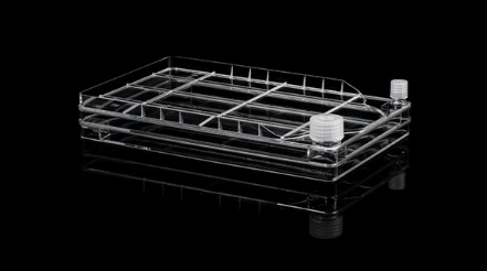
2 Layer Cell Factory One Wide Mouth And One Narrow Mouth
Facebook
X
Pinterest
Email
Huayi Cell Factory is a robust and easy-to-use cell culture platform for applications in the production of human and animal vaccines, therapeutic proteins, cell therapy, and gene therapy.
To address the needs of your workflow, We have three kinds of mouth, which is available on most cell culture products to ensure consistent performance from lot to lot and from format to format.
2 Layer Cell Factory One Wide Mouth And One Narrow Mouth
Huayi Cell Factory is a robust and easy-to-use cell culture platform for applications in the production of human and animal vaccines, therapeutic proteins, cell therapy, and gene therapy.
To address the needs of your workflow, We have three kinds of mouth, which is available on most cell culture products to ensure consistent performance from lot to lot and from format to format.
Our Blyscan™ Glycosaminoglycan Kit has been a ‘go-to’ Solution for reliable sGAG and Proteoglycan Analysis for many years! Blyscan utilises a dye-binding approach to quantitatively measure sulfated glycosaminoglycans (sGAG) and proteoglycans in cells, tissues and fluids from a wide range of in-vivo and in-vitro sources.
Colorimetric Detection (656nm) (Endpoint)
Understanding Glycosaminoglycans (GAGs) and Proteoglycans
Glycosaminoglycans (GAGs) are a type of negatively charged polysaccharide that play crucial roles in various biological processes. They are composed of repeated disaccharide units, typically of N-acetylated or N-sulfated hexosamine paired with a uronic acid (GlcA or IdoA) or galactose. Sulfate groups can also be added to give sulfated GAGs an overall negative charge that influences cell interactions and also enable binding by our Blyscan dye reagent.
Common examples of GAGs include Chondroitin Sulfate, Dermatan Sulfate, Heparin, Heparan Sulfate, and Keratan Sulfate. Note that Hyaluronic Acid is a non-sulfated GAG and cannot be detected by the Blyscan assay. If you need to measure hyaluronic acid instead, we recommend using our Purple-Jelley kit!
The Role of Glycosaminoglycans in Tissues
GAGs and proteoglycans have essential functions in tissues and organisms, providing biophysical support through scaffolding and maintaining cartilage hydration. They also play a vital role in biochemical processes such as cell adhesion and signalling.
What is the origin of the Blyscan assay name?
Blyscan is an Old English word meaning ‘to shine’ and from which the word ‘blush’, (blushing), may have been derived. This was an appropriate choice as the Blyscan Assay contains a blue dye which ‘blushes’ bright pink when it binds to sulphated glycosaminoglycans!
How does the Blyscan assay work?
Step 1. Blyscan dye reagent contains DMMB dye in an optimised buffer. Addition of Dye reagent to samples containing sGAG results in the formation of a dye/sGAG complex due to a charge interaction between dye and GAG sulfate groups.
Step 2. Over a 30 minute incubation Dye-labelled sGAGs precipitate out of solution and are collected by centrifugation. Following removal of unbound dye, the remaining bound dye is released from the complex by addition of dye dissociation reagent. Released dye is quantified spectrophotometrically.
Step 3. The sGAG content of unknown samples may be quantified by comparison against a calibration curve prepared using a standard of purified Chondroitin-4-sulfate supplied with the kit.
A list of suggested sample types can be found under the ‘Assay Specification‘ tab.
The Blyscan Dye reagent is formulated to miminise binding to other charged sample components such as nucleic acids, a problem with some older dye-based sGAG assays.
Assay range
2.5 – 50µg/ml
Limit of Detection
2.5µg/ml
Detection Method
Colorimetric Detection (656nm) (Endpoint)
Measurements per kit
110 in total (allows a maximum of 48 samples to be run in duplicate alongside a standard curve).
In-vivo: Liquid samples, including fluids such as urine, amniotic or synovial fluid.
In-vitro: Solid samples, such as deposited ECM on 2D/3D culture surfaces.by enzymatic treatment
In-vivo: Liquid samples, Culture media during 2D/3D cell culture.
The assay requires that sulfated polysaccahrides or sGAGs are in a soluble form. A preliminary enzymatic extraction step is required for solid samples (enzyme not supplied with kit).
The assay is not suitable for use with samples containing alginates or that comprise degraded sulfated disaccharide fragments.
Precautions
This kit is designed for research use only. Not for use in diagnostic procedures. Kit requires access to a centrifuge, as well as a spectrophotometer/colorimeter capable of absorbance detection at 656nm. Specific sample preparation protocols may require customer to provide further reagents, consult assay manual for further information.
Blyscan sGAG kit contents:
1. Blyscan Dye Reagent (1x110ml)
2.sGAG Reference Standard (1x5ml, 100µg/ml Bovine tracheal chondroitin 4-sulfate)
3. Dissociation Reagent (1x110ml)
4. Sodium Nitrite (1x15ml)
5. Acetic Acid (1x15ml)
6. Ammonium Sulfamate (1x15ml)
7. 1.5ml micro-centrifuge tubes for dye-labelling reaction.
8. Assay kit manual
NB: Additional reagents may be required for sample preparation prior to assay. Consult manual or contact us for further details.
Document
Our Blyscan™ Glycosaminoglycan Kit has been a ‘go-to’ Solution for reliable sGAG and Proteoglycan Analysis for many years! Blyscan utilises a dye-binding approach to quantitatively measure sulfated glycosaminoglycans (sGAG) and proteoglycans in cells, tissues and fluids from a wide range of in-vivo and in-vitro sources. Colorimetric Detection (656nm) (Endpoin
t-Boc-N-Amido-PEG7-propargyl is a crosslinking reagent that enables the formation of triazole linkage with azides under the catalyzation of copper. The Boc-protected amine can be deprotected under mild acidic conditions. By introducing PEG chain into the molecule, the hydrophilicity can be greatly improved. Reagent grade, for research purpose. Please contact us for GMP-grade inquiries.
Document
t-Boc-N-Amido-PEG7-propargyl is a crosslinking reagent that enables the formation of triazole linkage with azides under the catalyzation of copper. The Boc-protected amine can be deprotected under mild acidic conditions. By introducing PEG chain into the molecule, the hydrophilicity can be greatly improved. Reagent grade, for research purpose. Please contact us for GMP-grade inquiries.
The Multiplexing Index Primers contain primer mix for multiplexing library samples for Next Generation Sequencing (NGS) on the illumina platform. Multiplexing of NGS library samples will reduce sequencing costs by pooling multiple NGS libraries into a single flow cell lane.
With library multiplexing, unique index sequence is added to individual sample during NGS library preparation. Therefore, each DNA molecule can be identified after pooling of multiple samples based on the index information they have.
Each of our index primers contains a unique index sequence with 6 bases that can be used to identify libraries. Library multiplexing up to 48 samples is possible.
Multiplexing Index Primers (illumina platform): Even distribution of 48 samples using index primers. 48 libraries were made using the BioDynami NGS DNA Library Prep Kit (Cat. # 30009) and the BioDynami Multiplexing Index Primers (Cat. # 30072). Libraries were pooled at equal concentration and sequenced on the illumina HiSeq 2500. The numbers of reads from 48 libraries were analyzed.
List of index sequence for the primers (each of the index primer mix contains universal primer and one of the index primers). Index number and the index sequence are listed.
Sequence of the final library with index location:
Document
The Multiplexing Index Primers contain primer mix for multiplexing library samples for Next Generation Sequencing (NGS) on the illumina platform. Multiplexing of NGS library samples will reduce sequencing costs by pooling multiple NGS libraries into a single flow cell lane.