X96 Dual Ring Magnetic Plate 96-well with Integrated Cushion Base
Product Info
Document
Product Info
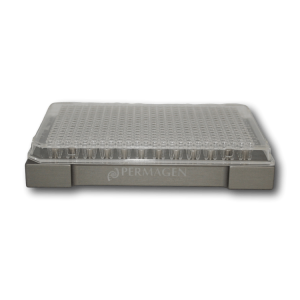
Permagen’s most universal ring magnet plate. From Low elution PCR applications up to 2 mL deep well, the X96 has you covered. No need to purchase two separate plates anymore. Smaller inner ring magnet allows for volumes as low as 5 µL (from PCR plates), larger outer magnet handles up to 2 mL deep well assays
ANSI/ SBS Footprint (127.75mm x 85.50mm) to fit into any automated liquid handling robot on bottom, SBS/ SLAS fit on top to accept any microplate
Integrated Cushion base for maximum recovery. Helps aid in set-up, robot positioning inconsistencies, and labware consumable differences
Features include solid aluminum alloy construction and hard coat anodized finish for years of trouble-free use, and compatible with any magnetic beads
Permagen’s most universal ring magnet plate. From Low elution PCR applications up to 2 mL deep well, the X96 has you covered. No need to purchase two separate plates anymore. Smaller inner ring magnet allows for volumes as low as 5 µL (from PCR plates), larger outer magnet handles up to 2 mL deep well assays
Basic Nucleic Acid DNA Amplification Kit 48 Reactions 20mins
Product Info
Document
Product Info
Product Description
Unlocking The Potential Of Nucleic Acid Amplification With DNA Amplification Kit (Basic)
Product Detail
Kit Storage and term of Validity
Storage term: stored at ≤-20℃,keep away from light, avoid heavy weight and repeated freezing and thawing.
Term of Validity: 14 months
Isothermal nucleic acid Principle Summary
The kit is based on rapid nucleic acid amplification technology at room temperature and constant temperature, its principle is that at room and constant temperature, the recombinase and primer form a protein/single-stranded nucleotide complex Rec/ssDNA, and invade the double-stranded DNA template with the help of auxiliary proteins and single-stranded binding protein SSB; then form a D-loop region at the invasion point and start to scan the DNA duplex, after finding the target region complementary to the primer and disintegration of the complex Rec/ssDNA, the polymerase also binds to the 3′ end of the primer to start the chain extension.
Isothermal nucleic acid Product Features
1/ High sensitivity and specificity, short reaction time.
2/ The reagent form is freeze-dried, stable and easy to operate.
3/ The reaction can be operated by metal bath and water bath pot without purchasing expensive PCR apparatus.
Technical Parameters:
Parameters
Details
Product Name
DNA Isothermal Amplification Kit Basic
Manufacturer
Amp-future
Storage Temperature
-20°C
Kit Components
Enzymes, Buffers ,Reagents
Packaging
48 Tests/box
Detection Limit
500-1000copies/µL
Shipping
ICE
Test Time
5-20mins
Isothermal nucleic acid Applications
Suitable for DNA isothermal rapid amplification kit(Basic type)
Primer: Require pair of nucleotide primers with the length of 25-35 bp.
DNA basic kit reaction temperature is 39 to 42℃ and time is 5-20 minutes.
Notes
1/ Please avoid nucleic acid contamination and set blank control during reaction due to the high sensitivity of the kit.
2/ Please take out the required quantity of MIRA reaction units for the experiment, and put the rest under storage conditions when performing the experiment.
Human Papillomavirus (HPV) 6/16 TaqMan PCR Detection Kits
Product Info
Document
Product Info
Overview
Detection kits for the HPV 6/16
Available in TaqMan format for analysis
More than 70 types of human papillomavirus (HPV) have been identified, and are generally classified as high-risk or low-risk depending on their relationship or lack of relationship with cancer and high-grade cervical intraepithelial neoplasia (CIN 2-3). HPV viruses are predominantly sexually transmitted and high-risk HPV types are a major risk factor for development of cervical cancer. HPV 16 has been considered as a high-risk cancer associated HPV type. The low-risk HPV type 6 has been associated with the presence of genital warts. There are many other low-risk HPV types that are not associated with genital warts or cervical cancer. Until now, HPV cannot be cultured in vitro, and immunological tests are inadequate to determine the presence of HPV cervical infection. On the other hand, biopsies can be analyzed by nucleic acid hybridization to directly detect the presence of HPV DNA.
HPV 6/16 TaqMan PCR Kit, 100 reactions
Ready to use format, including Master Mix for the target and PCR control to monitor for PCR inhibition and validate the quality
Specific Primer and Probe mix for the pathogen/virus/viroid of interest
Primer and Probe mix
Positive and negative control to confirm the integrity of the kit reagents
HPV 6/16 TaqMan PCR Probe/Primer Set and Controls, 100 reactions
Specific Primer/Probe mix and Positive Control for the pathogen/virus/viroid of interest
Nuclease-free water
Can be used together with Norgen’s PCR Master Mix (#28007) or customer supplied master mix
For research use only and NOT intended for in vitro diagnostics.
Storage Conditions and Product Stability All kit components can be stored for 2 years after the date of production without showing any reduction in performance.
All kit components should be stored at -20°C upon arrival.
Component
TaqMan Probe Cat. TM42050 (100 preps)
TaqMan Probe/Primer and Control Set Cat. TM42010 (100 preps)