
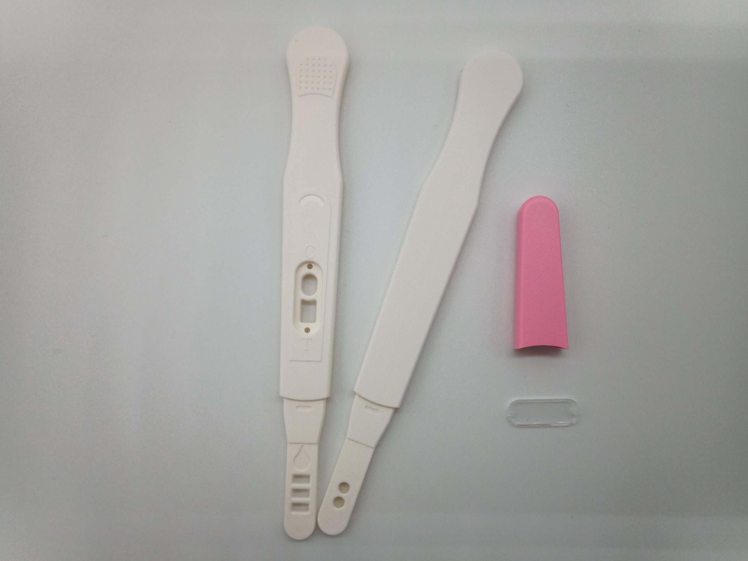
Description
- • Appearance: White/Pink with lens
- • Length of strip: 86mm
- • Width of strip: 4.5mm
- • Length of device: 136mm
- • Width of device: 16mm


Norgen’s Plant/Fungi Total RNA Purification Kit provides a rapid method for the isolation and purification of total RNA, including virus and viroid RNA, from a wide range of plants. Total RNA can be purified from fresh or frozen plant tissues, plant cells or filamentous fungi samples using this kit. All sizes of RNA are purified, including microRNA (miRNA) . The procedure is rapid and convenient.
The RNA is purified without the use of phenol or chloroform. The purified RNA is of the highest quality, and can be used in a number of downstream applications including real time PCR, reverse transcription PCR, Northern blotting, RNase protection and primer extension, and expression array assays.
Norgen’s Plant/Fungi Total RNA Purification Kit is also available in a 96-well (High Throughput) format for high throughput applications. Purification with the 96-well plates can be performed using either a vacuum manifold or centrifugation.
Figure 1 / 4

Click for expanded view
| Kit Specifications – Spin Column | |
| Maximum Column Binding Capacity | 50 μg |
| Maximum Column Loading Volume | 650 μL |
| Size of RNA Purified | All sizes, including small RNA (< 200 nt) |
| Maximum Amount of Starting Material: Plant Tissues Plant Cells Fungi | 50 mg 1 x 10⁶ cells 50 mg (wet weight) |
| Average Yield* 50 mg Tomato Leaves 50 mg Tobacco Leaves 50 mg Plum Leaves 50 mg Grape Leaves 50 mg Peach Leaves | 60 μg 60 μg 32 μg 35 μg 30 μg |
| Time to Complete 10 Purifications | 30 minutes |
* Yield will vary depending on the type of sample processed.
* Yield will vary depending on the type of sample processed.
Storage Conditions and Product Stability
All solutions should be kept tightly sealed and stored at room temperature. This kit is stable for 2 years after the date of shipment.
Tobacco (Nicotiana tabacum)
Tomato (Lycopersicon esculentum)
Pepper (Capsicum annuum)
Potato (Solanum tuberosum)
Arabidopsis thaliana1
Peach (Prunus persica)
Apple (Malus sp.)
Pear (Pyrus sp.)
Grape vine (Vitis sp.)
Plum (Prunus sp.)
Palm (Arecaceae)
Pine needle (Pinaceae)
Strawberry
Raspberry
Blackberry
Herbs
Persimmon (Ebenaceae)
Potato tuber (Solanum)
Plum fruit
Citrus
Vanilla bean
Cotton (Gossypium)
Mangrove
Chrysanthemum
Grape berry skin
Kiwi leaves
Peach (fruits and flowers)
Soy bean (legume)
Eastern White Red Cedar
Corn leaves
Cucumber leaves
Aspergillus niger
Mucor racemosus
Cladosporium cladosporioides
Fusarium oxysporum
Penicillium sp.
Botrytis cinerea (Botryotinia fuckeliana)
Pichia sp.
Rhizopus oryzae
Alternaria tenuissima
| Component | Cat. 25800 (50 preps) | Cat. 31350 (100 preps) | Cat. 25850 (250 preps) | Cat. 31900 (192 preps) |
|---|---|---|---|---|
| Lysis Buffer C | 60 mL | 1 x 30 mL, 1 x 60 mL | 3 x 60 mL | 2 x 60 mL |
| Wash Solution A | 38 mL | 38 mL | 1 x 18 mL, 2 x 38 mL | 2 x 38 mL |
| Elution Solution A | 6 mL | 6 mL | 20 mL | 20 mL |
| Filter Columns | 50 | 100 | 250 | – |
| Spin Columns | 50 | 100 | 250 | – |
| 96-Well Plate | – | – | – | 2 |
| Adhesive Tape | – | – | – | 4 |
| Collection Tubes | 100 | 200 | 500 | – |
| 96-Well Collection Plate | – | – | – | 2 |
| Elution Tubes (1.7 mL) | 50 | 100 | 250 | – |
| 96-Well Elution Plate | – | – | – | 2 |
| Product Insert | 1 | 1 | 1 | 1 |
The BK virus is a member of the polyomavirus family. BK viral infections are typically asymptomatic in healthy individuals, however very mild symptoms may appear including mild respiratory infections and fever. Once an individual has been infected the virus disseminates to the kidneys and the urinary tract where it remains for the lifetime of the individual. Infections with BK virus in immunocompromised or immunosupressed patients are much more severe and may involve renal dysfunction. In fact, in kidney transplant patients the immunosupressive drugs required for the transplant may allow the virus to replicate within the graft, resulting in a disease called BK virus nephropathy (BKVN). It is thought that 1-10% of renal transplant patients progress to BK virus nephropathy (BKVN) and up to 80% of these patients are reported to have lost their grafts. The onset of nephritis can occur as early as several days post-transplant to as late as 5 years. The mode of transmission of the virus is not clear, however it has been suggested that BKV may be transmitted through respiratory fluids or urine, since infected individuals periodically excrete virus in the urine. This virus can be diagnosed by BKV blood & urine testing, in addition to carrying out a biopsy in the kidneys. PCR techniques are now widely used to identify the virus.
BKV TaqMan PCR Kit, 100 reactions
BKV TaqMan PCR Probe/Primer Set and Controls, 100 reactions
For research use only and NOT intended for in vitro diagnostics.
Figure 1 / 3
Click for expanded view
Storage Conditions and Product Stability
All kit components can be stored for 2 years after the date of production without showing any reduction in performance.
All kit components should be stored at -20°C upon arrival. Repeated thawing and freezing (> 2 x) of the Master Mix and Positive Control should be avoided, as this may affect the performance of the assay. If the reagents are to be used only intermittently, they should be frozen in aliquots.
| Component | Cat. TM36550 (100 preps) | Cat. TM36510 (100 preps) |
|---|---|---|
| MDx TaqMan 2X PCR Master Mix | 2 x 700 μL | – |
| BKV Primer & Probe Mix | 280 μL | 280 μL |
| BKV Positive Control | 150 μL | 150 μL |
| Nuclease-Free Water (Negative Control) | 1.25 mL | 1.25 mL |
| Product Insert | 1 | 1 |

Propargyl-PEG13-alcohol can reacts with azide-bearing compounds or biomolecules via copper catalyzed Click Chemistry to form stable triazole linkage. The hydrophilic PEG units increase the water-solubility of the molecule. Reagent grade, for research purpose. Please contact us for GMP-grade inquiries.

Propargyl-PEG13-alcohol can reacts with azide-bearing compounds or biomolecules via copper catalyzed Click Chemistry to form stable triazole linkage. The hydrophilic PEG units increase the water-solubility of the molecule. Reagent grade, for research purpose. Please contact us for GMP-grade inquiries.