This product provides fast and easy methods for purification of total DNA for reliable PCR and Southern blotting. Total DNA(e.g., genomic, viral, mitochondrial) can be purified from small volume of blood, tissue and dry blood spots.
Detail
Introduction
This product provides fast and easy methods for purification of total DNA for reliable PCR and Southern blotting. Total DNA(e.g., genomic, viral, mitochondrial) can be purified from small volume of blood, tissue and dry blood spots.
Details
Specifications
Features
Specifications
Main Functions
Isolation total DNA from 1-10μl blood, <10mg tissue, urine, blood stain, seminal stain
Applications
PCR, southern bolt and virus detection, etc.
Purification method
Mini spin column
Purification technology
Silica technology
Process method
Manual (centrifugation or vacuum)
Sample type
Animal tissues, blood stain, urine, seminal stain and various forensic samples
Sample amount
Blood:1-100μl, Tissue:<10mg
Elution volume
Time per run
Liquid carrying volume per column
Binding yield of column
Principle
This product is based on silica Column purification. The sample is lysed and digested with lysate and protease, DNA is released into the lysate. Transfer to an adsorption column. Nucleic acid is adsorbed on the membrane, while protein is not adsorbed and is removed with filtration. After washing proteins and other impurities, nucleic acid was finally eluted with low-salt buffer (10mmTris, pH9.0, 0.5mm EDTA).
Advantages
Fast – several samples can be extracted in 20 minutes (after digestion)
High purity – purified DNA can be directly used in various downstream applications
High recovery – DNA can be recovered at the level of PG
Good repeatability – silica technology can obtain ideal results every time
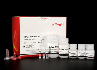
Kit Contents
Contents
D312502
D312503
Purification Times
50 Preps
250 Preps
Buffer ATL
15 ml
60 ml
Buffer AL
15 ml
60 ml
Buffer GW1*
22 ml
66 ml
Buffer GW2*
20 ml
2 x 50 ml
Carrier RNA
310 μg
2 x 310 µg
Proteinase K
24 mg
120 mg
Protease Dissolve Buffer
1.8 ml
10 ml
Buffer AE
15 ml
60 ml
HiPure DNA Mini Columns I
50
2 x 125
2 ml Collection Tubes
100
5 x 100
Storage and Stability
Carrier RNA and Proteinase K should be stored at 2–8°C upon arrival. However, short-term storage (up to 12 weeks) at room temperature (15–25°C) does not affect its performance. The remaining kit components can be stored dry at room temperature (15–25°C) and are stable for at least 18 months under theseconditions.
Experiment Data
Other Products
Sircol-2.0 Soluble Collagen assay kit
Product Info
Document
Product Info
What is Collagen?
Collagen is a fundamental component of the extracellular matrix, and the predominant protein in animals, constituting around 30% of total protein mass. A glycoprotein, it is well known for its triple helical structure. This is formed from three polypeptide α-chains with Gly-X-Y repeating residues (Gly for Glycine, X for proline, and Y for hydroxyproline).
Types of Collagen
Over 28 types of collagens have been identified, with Type I collagen being the most abundant. It’s prevalent in ligaments, tendons, skin, and bone tissue. Its mature, insoluble form grants it remarkable strength, making it vital for the mobility of organisms. Collagen also has biochemical functions, influencing cell growth, proliferation, and differentiation.
This version of the kit is designed to detect and measure SOLUBLE forms of collagen. Chose the Sircol Insoluble collagen kit if you need to analyse INSOLUBLE collagen.
Applications of Collagen
Collagen, with its diverse properties, finds utility in various industries. It plays a role in medicine for wound healing and has an expanding role in tissue engineering and cell culture for biomedical purposes. It’s gaining popularity in the cosmetic industry for skin rejuvenation and is used in chemical formulations and the food industry as a functional food supplement and additive.
How does Sircol 2.0 detect collagen?
The Sircol 2.0 dye reagent includes Sirius Red, a linear anionic dye with sulfonic acid side chains. This reagent is specially formulated to bind to the Gly-X-Yn helical structure of soluble collagen under assay conditions.
*The improved formulation of Sircol 2.0 dye enables a greater degree of dye-collagen specificity (compared to our previous S1000 assay kit).
Overview of the Sircol 2.0 assay process:
Step 1. Prepared samples are placed in the wells of the assay microplate, together with Sircol Dye Reagent. After 30 minutes mixing, any collagen-dye complexes will form as a precipitate. These are collected on the base of the microplate wells by centrifugation.
Step 2. Unbound dye is removed by gentle aspiration, followed by a rinse with Plate Wash Reagent.
Step 3. Following further centrifugation, collagen-bound dye is eluted by incubation with a Dye Release Reagent. Eluted dye is detected ‘in-situ’ by spectrophotometric analysis of the microplate at 556nm.
Step 4. The collagen content of unknown samples can be quantified by comparison against a calibration curve, prepared using the Collagen Reference Standard supplied with the kit.
A list of suggested sample types can be found under the ‘Assay Specification‘ tab.
Colorimetric Detection (556nm) (Endpoint), Requires a microplate centrifuge.
Measurements per kit
96 in total (allows a maximum of 41 samples to be run in duplicate alongside a standard curve).
Suitable Samples
Soluble* collagens from mammalian**:
In-vivo: Tissues, cartilages and fluids.
In-vitro: Extracellular matrices / Conditioned media from 2D/3D culture environments.
The straightforward sample processing and analysis of Sirco 2.0 make it a good alternative to conventional hydroxyproline analysis.
*Prior salt/acid/acid-pepsin extraction may be necessary to release soluble collagen.
**Sircol 2.0 is primarily designed for use with in-vivo / in-vitro samples of mammalian origin. Collagens originating from other taxonomic groups and kingdoms can also be analysed. See note on p6 of manual for further information.
Precautions
This kit is designed for research use only. Not for use in diagnostic procedures. Kit requires access to a microplate centrifuge* (see note below), as well as a spectrophotometer/colorimeter capable of absorbance detection at 556nm. Specific sample preparation protocols may require customer to provide further reagents, consult assay manual for further information.
*As a minimum, we recommend that the centrifuge can centrifuge a 96-well microplate at 400 x g for 120 minutes. Higher speed centrifuges are recommended (up to a maximum of 2000 x g), allowing a reduction in centrifuge time.
Sircol 2.0 kit contents:
1. Dye Reagent (1x20ml)
2. Collagen Reference Standard (1x5ml, 200µg/ml of soluble Bovine collagen)
3. Plate Wash Reagent (1x28ml)
4. Collagen Concentration Reagent (1x25ml)
5. Neutralisation Reagent (1x8ml)
6. Dye Release Reagent (1x25ml)
7. Assay Microplate (1×96-wells)
8. Microplate Seals (6x)
9. Documentation (QuickStart Guide / Manual / Certificate of Analysis)
NB: Additional reagents may be required for sample preparation prior to assay. Consult manual or contact us for further details. This kit requires the use of a microplate centrifuge, capable of centrifuging a 96-well microplate at 400 x g for 120 minutes. Higher speed centrifuges are recommended (up to a maximum of 2000 x g), allowing a reduction in centrifuge time.
Document
Experience user-friendly detection & measurement of Soluble Collagen with Sircol™ 2.0! Our latest kit simplifies collagen quantification within in-vivo / in-vitro samples. Sircol 2.0 offers enhanced sensitivity and accuracy compared to our previous Sircol kit.
An enhanced PCR master mix for allele-specific assays. Improved signal to noise ratio and tight clustering. Developed specifically for genotyping direct from crude DNA samples.
PACE 2.0 Genotyping Master Mix ensures an unrivalled signal-to-noise ratio and produces tight data clusters, even when working with high-throughput, crude DNA preps, resulting in consistently exceptional performance. Efficiently streamline your workflow and reduce costs without compromising the quality of your results.
PACE 2.0 Genotyping Master Mix is an ideal solution for challenging starting material. PACE 2.0 has been specially formulated to overcome the obstacles presented by common PCR inhibitor compounds, such as phenols and tannins. Even notoriously tricky samples like oil palm and conifers can still be assayed using hot shot or other crude DNA prep methods and deliver reliable and accurate data.
PACE 2.0 Genotyping Master Mix uses a novel, universal, fluorescent reporting cassette to produce machine-readable fluorescent signals corresponding to genotypes. PACE 2.0 compatible genotyping assays are comprised of two competitive allele-specific forward primers (which differ in their terminal 3’ bases and unique 5’ tail sequences) and a common, reverse primer. PACE 2.0 Genotyping Master Mix is supplied with ROX normalising dye at a range of levels to ensure compatibility with your qPCR instrument or reader.
Genotyping assay designs are available from 3CR Bioscience through our free assay-design service; once designed, users can purchase assay primers independently or through 3CR Bioscience using our partial or full-assay validation service. PACE 2.0 Genotyping Master Mix is also compatible with KASP™ and Amplifluor® marker assays.
REQUIRED COMPONENTS
qPCR machine or Thermocycler + Fluorescent plate reader
PCR plate or equivalent and appropriate optically clear seal
Real-time fluorescent DNA amplification made possible with a TwistAmp® exo kit. Recommended for users who want to combine TwistDx’s RPA amplification technology with the use of TwistDx’s proprietary fluorescent TwistAmp® exo probe in a homogenous format. The user need only supply primers, probe, and template. See manual for more information. Click to order oligonucleotides.
Document
Real-time fluorescent DNA amplification made possible with a TwistAmp® exo kit. Recommended for users who want to combine TwistDx’s RPA amplification technology with the use of TwistDx’s proprietary fluorescent TwistAmp® exo probe in a homogenous format. The user need only supply primers, probe, and template. See manual for more information. Click to order oligonucleotides.