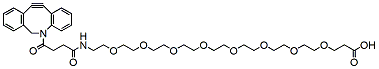
DBCO-PEG8-acid is an analog of DBCO-Acid with hydrophilic PEG linker and a DBCO group. The DBCO groups is commonly used for Click Chemistry reactions. The hydrophilic PEG chain allows for increased water solubility of compounds in aqueous media. The terminal carboxylic acid can react with primary amine groups in the presence of activators (e.g. EDC, or HATU) to form a stable amide bond. Reagent grade, for research purpose. Please contact us for GMP-grade inquiries.
Detail
DBCO-PEG8-acid is an analog of DBCO-Acid with hydrophilic PEG linker and a DBCO group. The DBCO groups is commonly used for Click Chemistry reactions. The hydrophilic PEG chain allows for increased water solubility of compounds in aqueous media. The terminal carboxylic acid can react with primary amine groups in the presence of activators (e.g. EDC, or HATU) to form a stable amide bond. Reagent grade, for research purpose. Please contact us for GMP-grade inquiries.
Other Products
[DS1001] FluoroStain™ DNA Fluorescent Staining Dye (Green, 10,000X), 500 μl x 5
Product Info
Document
Product Info
Description
The FluoroStain™ DNA Fluorescent Staining Dye is designed to be a safer replacement for conventional Ethidium bromide (EtBr) which poses a significant health and safety hazard for its users. The FluoroStain™ DNA Fluorescent Staining Dye offers at least 10 times sensitivity in DNA detection levels, and is capable of detecting double stranded DNA (dsDNA) fragments up to 0.04 ng in electrophoresis analysis. The FluoroStain™ DNA Fluorescent Staining Dye shows a high specificity to the dsDNA, with negligible background signal, making the destaining process entirely optional. FluoroStain™ DNA Fluorescent Staining Dye is compatible with both the conventional ultra violet gel-illuminating systems as well as the less harmful long wave length blue light illumination systems. The emission when bound to dsDNA is 522 nm, while its excitation peaks are at 270, 370 and 497 nm.
Features:
Excellent for post staining
Sensitivity: 0.04 ng DNA
A safer alternative to EtBr
Compatibility: suitable to blue or UV light
Increased cloning efficiency (blue light)
Storage
Protected from light 4°C for 12 months -20°C for 24 months
Document
The FluoroStain™ DNA Fluorescent Staining Dye is designed to be a safer replacement for conventional Ethidium bromide (EtBr) which poses a significant health and safety hazard for its users. The FluoroStain™ DNA Fluorescent Staining Dye offers at least 10 times sensitivity in DNA detection levels, and is capable of detecting double stranded DNA (dsDNA) fragments up to 0.04 ng in electrophoresis analysis. The FluoroStain™ DNA Fluorescent Staining Dye shows a high specificity to the dsDNA, with negligible background signal, making the destaining process entirely optional. FluoroStain™ DNA Fluorescent Staining Dye is compatible with both the conventional ultra violet gel-illuminating systems as well as the less harmful long wave length blue light illumination systems. The emission when bound to dsDNA is 522 nm, while its excitation peaks are at 270, 370 and 497 nm.
High Efficiency RNA Isothermal PCR Kit Stable Easy Operate
Product Info
Document
Product Info
Product Description
High-Efficiency Isothermal RNA Amplification Kit, Stable And Easy To Operate
Product Detail
Kit Storage and term of Validity
Storage term: stored at ≤-20℃,keep away from light, avoid heavy weight and repeated freezing and thawing.
Term of Validity: 14 months
Isothermal nucleic acid Principle Summary
This kit is based on a rapid nucleic acid amplification technology at room temperature and constant temperature:at room temperature and constant temperature(generally 39℃~42℃),reverse transcriptaseuses specific primers and template RNA tosynthesize cDNA strands,and binds the auxiliary protein and single strand with the help of the protein, the recombinase and the primer form a complex; perform a homology search and bind the target homology domain, at this time a D-loop region is formed at the homology positionand strand exchange begins; accompanied by the recombinase from the complex upon dissociation,the polymerase also bindsto the 3′ end of the primer,initiating chainextension.It is suitable for laboratory-level RNA amplification and RNA amplification for other detection purposes.
Isothermal nucleic acid Product Features
1/ High sensitivity and specificity, short reaction time.
2/ The reagent form is freeze-dried, stable and easy to operate.
3/ The reaction can be operated by metal bath and water bath pot without purchasing expensive PCR apparatus.
Technical Parameters:
Parameters
Details
Product Name
RNA Isothermal Amplification Kit Basic
Manufacturer
Amp-future
Storage Temperature
-20°C
Kit Components
Enzymes, Buffers ,Reagents
Packaging
48 Tests/box
Detection Limit
500-1000copies/µL
Shipping
ICE
Test Time
5-20mins
Isothermal nucleic acid Applications
Suitable for RNA isothermal rapid amplification kit(Basic type)
Primer: Require pair of nucleotide primers with the length of 25-35 bp.
RNA basic kit reaction temperature is 39 to 42℃ and time is 5-20 minutes.
Notes
1/ Please avoid nucleic acid contamination and set blank control during reaction due to the high sensitivity of the kit.
2/ Please take out the required quantity of MIRA reaction units for the experiment, and put the rest under storage conditions when performing the experiment.