

DD5 Low Speed 5000rpm Swing rotor 4x800ml lab Centrifuge Adapters for 5/10/15/50ml tubes.

DD5 Low Speed Large Capacity Centrifuge Application:
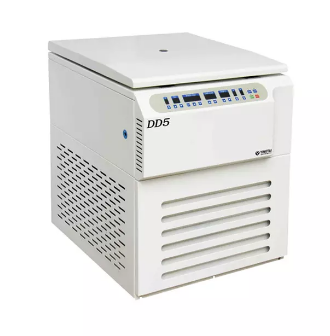
DD5 Lab centrifuge widely used in the fields of pharmaceutical factory and laboratory, can hold swing out rotors and angle rotors both.
DD5 Low Speed Large Capacity Centrifuge Features:
1. Brushless frequency motor which in great torque, free maintenance, no powder pollution, quick in speed up and down.
2. Digital display which indicates the program, speed, time, RCF and temperature.
3. Micro computer control, there are 10 kinds of program and 10 kinds of acceleration/deceleration for your choice.
4. There are kinds of rotors for your choice.
5. Electric lid lock, compact design, super speed and imbalance protection.
6. The centrifuge body is made of high-quality steel, safe and reliable.
DD5 Low Speed Laboratory Centrifuge Technical Parameters:
| Max. Speed | 5000rpm |
| Max.RCF | 4730xg |
| Max.Capacity | 4x800ml |
| Timer | 0~99mins |
| RPM/RCF Convert | Yes |
| Noise(DB) | ≤58 |
| Acc/Dec | 10 Rates |
| Speed Accuracy | ±20rpm |
| Voltage(V/HZ) | AC 220V/110V 50HZ/60HZ |
| Dimension(LxWxH mm) | 720x540x830 |
| Net Weight(Kg) | 108kg |
| Certificates | CE,ISO |
| Warranty | 1 Year |
DD5 Low Speed Laboratory Centrifuge Matched Rotors:
| Order No. | Rotor type | Max.Speed (rpm) | Max.Volume (ml) | Max.RCF (xg) |
| No30671 | Swing out rotor | 4,000 | 4x800ml(round cup) | 3,450 |
| No30679 | Swing out rotor | 4,000 | 4x500ml(square cup) | 3,450 |
| No30696 | Swing out rotor | 4,000 | 4x500ml(round cup) | 3,530 |
| No31377 | Swing out rotor | 5,000 | 4x50ml | 4,730 |
| 4x100ml | 4,730 | |||
| No30592 | Swing out rotor | 4,000 | 4x30x7ml | 3,140 |
| 4x40x7ml | 3,270 | |||
| 4x18x10ml | 3,140 | |||
| No31491 | Mircoplater rotor | 4,000 | 2x4x96well | 2,910 |
| No31494 | Microplater rotor | 4,000 | 4x4x96well | 2,940 |
| No30627 | Angle rotor | 5,000 | 30x15ml | 3,830 |
| No30641 | Angle rotor | 5,000 | 12x50ml | 3,860 |
| No30642 | Angle rotor | 4,000 | 24x50ml | 2,970 |
| No30614 | Angle rotor | 5,000 | 6x100ml | 3,130 |
| No30643 | Angle rotor | 4,000 | 12x100ml | 2,970 |
The Corners of our Company____________________________________


