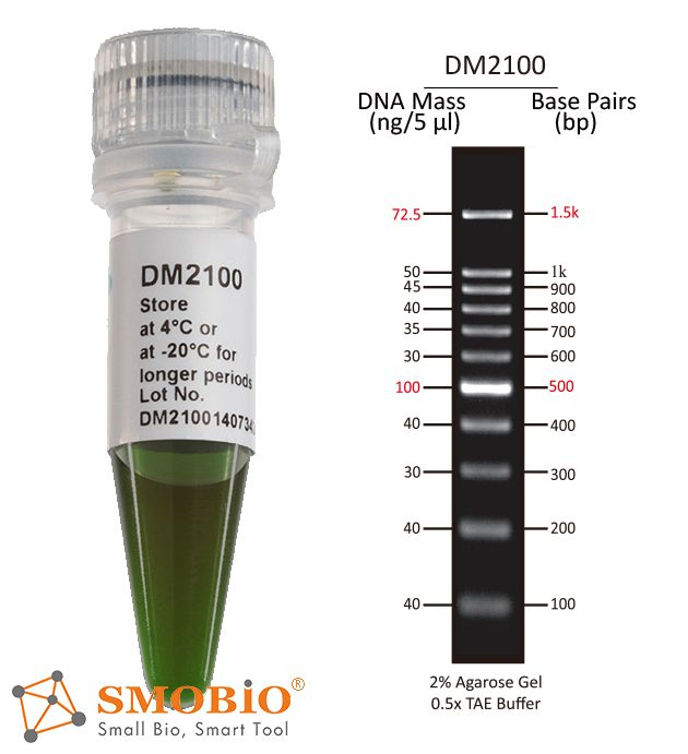
The DM2100 ExcelBand™ 100 bp DNA Ladder is a ready-to-use DNA ladder, which is pre-mixed with loading dye for direct gel loading. The DNA ladder DM2100 is composed of 11 individual DNA fragments: 1.5k, 1k, 900, 800, 700, 600, 500, 400, 300, 200 and 100 bp derived from a mixture of PCR products and specifically digested plasmid DNA. This product contains two enhanced bands (1.5 kb and 500 bp) for easier reference. In addition, two tracking dyes, Xylene cyanol FF and Orange G which mimic the migration of 4,000 bp and 50 bp dsDNA during electrophoresis are also added for real time monitoring.
Detail
Description
The DM2100 ExcelBand™ 100 bp DNA Ladder is a ready-to-use DNA ladder, which is pre-mixed with loading dye for direct gel loading. The DNA ladder DM2100 is composed of 11 individual DNA fragments: 1.5k, 1k, 900, 800, 700, 600, 500, 400, 300, 200 and 100 bp derived from a mixture of PCR products and specifically digested plasmid DNA. This product contains two enhanced bands (1.5 kb and 500 bp) for easier reference. In addition, two tracking dyes, Xylene cyanol FF and Orange G which mimic the migration of 4,000 bp and 50 bp dsDNA during electrophoresis are also added for real time monitoring.
Features
Sharp bands
Quick reference— enhanced bands
Ready-to-use— premixed with loading dye for direct loading
Stable— room temperature storage over 6 months
Source
Phenol extracted PCR products and dsDNA digested with specific restriction enzymes, equilibrated in 10 mM Tris-HCl (pH 8.0) and 10 mM EDTA.
Range
100 ~ 1,500 bp
Concentration
52.2 µg/ 500 µl
Recommended loading volume
5 µl/ well
Storage
Room temperature for 6 months 4°C for 12 months -20°C for 36 months
Other Products
[PS1000] FluoroStain™ Protein Fluorescent Staining Dye (Red, 1,000X), 1 ml
Product Info
Document
Product Info
Description
The FluoroStain™ Protein Fluorescent Staining Dye (Red, 1000×) is designed to substitute the common Coomassie Blue protein staining method, offering greater sensitivity and ease of operation. Unlike Coomassie Blue stain, the FluoroStain Protein Fluorescent Staining Dye binds to protein with high specificity, making destaining process an option rather than a requirement. With further reduction of background signals via destaining process, the FluoroStain™ is capable of achieving detection level parallel to silver staining without specialized imaging equipment, making it one of the most sensitive dyes available. In addition to its remarkable sensitivity, the FluoroStain™ Protein Fluorescent Staining Dye (Red, 1000×) brings a more reliable and safer user experience, since the stained gel can be visualized with blue-light illumination, avoiding the risk of skin/ eye damage caused by UV light. For best result, we suggest using B-BOX™ Blue Light LED epi-illuminator to visualize and analyze the gel stained with FluoroStain Protein Fluorescent Staining Dye (Red, 1000×). The FluoroStain™ Protein Fluorescent Staining Dye is compatible to the analysis of mass spectra, i.e. LC-MS/MS, MALDI-TOF, etc.
Spectral Characteristics
When it is bound with bovine serum albumin (BSA), the fluorescent emission of FluoroStain Protein Fluorescent Staining Dye can be excited by UV and blue light sources, with excitation peaks around 369 and 517 nm and emission at 605 nm. In absence of BSA, FluoroStain Protein Fluorescent Staining Dye shows ignorable fluorescence as compared with protein-bound form, therefore giving a clear background for photographic analysis.
These spectral characteristics made this fluorescent dye compatible with a wide variety of gel reading facilities, including UV/ blue light epi- and transilluminator, argon laser and mercury-arc lamp excitation gel scanners.
Storage
Protected from light -20°C for 24 months
Document
The FluoroStain™ Protein Fluorescent Staining Dye (Red, 1000×) is designed to substitute the common Coomassie Blue protein staining method, offering greater sensitivity and ease of operation. Unlike Coomassie Blue stain, the FluoroStain Protein Fluorescent Staining Dye binds to protein with high specificity, making destaining process an option rather than a requirement. With further reduction of background signals via destaining process, the FluoroStain™ is capable of achieving detection level parallel to silver staining without specialized imaging equipment, making it one of the most sensitive dyes available. In addition to its remarkable sensitivity, the FluoroStain™ Protein Fluorescent Staining Dye (Red, 1000×) brings a more reliable and safer user experience, since the stained gel can be visualized with blue-light illumination, avoiding the risk of skin/ eye damage caused by UV light. For best result, we suggest using B-BOX™ Blue Light LED epi-illuminator to visualize and analyze the gel stained with FluoroStain Protein Fluorescent Staining Dye (Red, 1000×). The FluoroStain™ Protein Fluorescent Staining Dye is compatible to the analysis of mass spectra, i.e. LC-MS/MS, MALDI-TOF, etc.
Not all cyanobacterial strains produce toxins. However, the toxin-producing strains cannot be distinguished from the nontoxin-producing strains by traditional light microscopy, commonlyused to monitor water bodies. An alternative for the differentiation of potentially toxic strains from nontoxic strains is to use molecular methods to detect the presence of toxin biosynthetic genes. Such methods are already available and could be used for the detection and identification of potential microcystin and nodularin producers present in environmental samples (Attogene catalog number NA2024).
Screening for the toxin itself, can be very costly. In turn, real time PCR for the detection of a gene region responsible for assembling in cyanobacterial strains and environmental samples can be a key indicator for the prescense of cyanobacteria capable of expressing the aetokthonotoxin toxin. Attogen has thus, designed primer pairs and probes targeting a the conserved gene region in order to enable the amplification and detection of several producer genera using real time PCR. Screening for the toxin genes can save significant costs and act as a triage for samples needing to be analyzed for the toxin itself.
Cyanobacterial neurotoxin aetokthonotoxin (AETX), a peculiar pentabrominated biindole alkaloid implicated in fatal Vacuolar Myelinopathy. This neurodegenerative disease was first recorded in 1994 during an outbreak of bald-eagle poisonings at De Gray Lake in Arkansas, USA. AETX was experimentally confirmed to be produced by the true branching heterocytous cyanobacterium Aetokthonos hydrillicola. The production of AETX is dependent on bromide (Br−) availability, and likely linked to its hyper-accumulation by the host plan. Thus regular monitoring of A. hydrillicola (accompanied by assessment of Br− and AETX levels) is highly advisable to predict the possible threat of further VM outbreaks.
The cyanobacterial AetA gene which encodes the unique FAD-dependent halogenase involved in the pathway for AETX synthesis has been adapted to develop a -aetokthonotoxin specific quantitative PCR (qPCR) assay.
Document
Real time qPCR kit for AetA gene For screening aetokthonotoxin gene cluster Use in combination with Attogene Algae DNA isolation kit