

Description
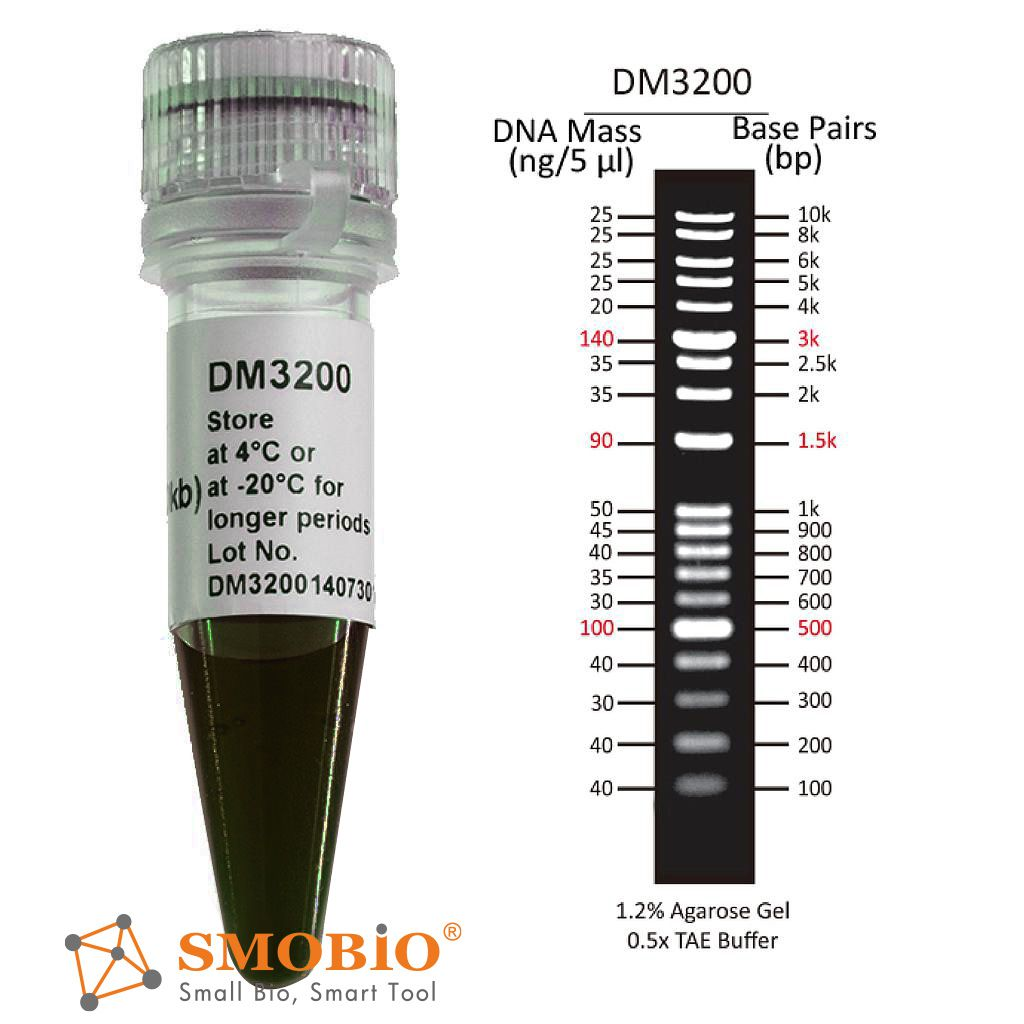
The DM3200 ExcelBand™ 1 KB Plus (0.1-10 kb) DNA Ladder is a ready-to-use DNA ladder, which is pre-mixed with loading dye for direct gel loading. The DNA Ladder DM3200 is composed of 19 individual DNA fragments: 10k, 8k, 6k, 5k, 4k, 3k, 2.5k, 2k, 1.5k, 1k, 900, 800, 700, 600, 500, 400, 300, 200, and 100 bp derived from a mixture of PCR products and specifically digested plasmid DNA. This product contains three enhanced bands (3 kb, 1.5 kb and 500 bp) for easier reference. In addition, three tracking dyes, Xylene cyanol FF, Bromophenol blue and Orange G which mimic the migration of 4,000 bp, 500 bp and 50 bp dsDNA during electrophoresis are also added for real time monitoring.
Features
- Sharp bands
- Quick reference— enhanced bands
- Ready-to-use— premixed with loading dye for direct loading
- Stable— room temperature storage over 6 months
Source
Phenol extracted PCR products and dsDNA digested with specific restriction enzymes, equilibrated in 10 mM Tris-HCl (pH 8.0) and 10 mM EDTA.
Range
100 ~ 10,000 bp
Concentration
87 µg/ 500 µl
Recommended loading volume
5 µl/ well
Storage
Room temperature for 6 months
4°C for 12 months
-20°C for 36 months


