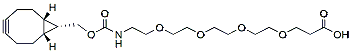
endo-BCN-PEG4-acid is a click chemistry reagent with a BCN group witha terminal carboxylic acid (CO2H). The terminal carboxylic acid is reactive with primary amine groups in the presence of activators (e.g. EDC, or HATU) to form a stable amide bond. The BCN group can react with azide -tagged compound or biomolecules. The hydrophilic PEG spacer increases solubility in aqueous media. Reagent grade, for research purpose. Please contact us for GMP-grade inquiries.
Detail
endo-BCN-PEG4-acid is a click chemistry reagent with a BCN group witha terminal carboxylic acid (CO2H). The terminal carboxylic acid is reactive with primary amine groups in the presence of activators (e.g. EDC, or HATU) to form a stable amide bond. The BCN group can react with azide -tagged compound or biomolecules. The hydrophilic PEG spacer increases solubility in aqueous media. Reagent grade, for research purpose. Please contact us for GMP-grade inquiries.
Other Products
Plasmid MiniPrep Kit & High Throughput Kit (Magnetic Beads)
Product Info
Document
Product Info
Plasmid MiniPrep Kit & High Throughput Kit (Magnetic Beads)
The kits were developed for plasmid miniprep directly from bacterial cultures. 100% centrifuge-free; NO vacuum needed; No column needed.
Plasmid MiniPrep Kit (Magnetic Beads), Cat.# 50012 – Use 2 ml of bacteria culture directly
Plasmid MiniPrep High Throughput Kit (Magnetic Beads), Cat.# 50011 – High throughput: Use 0.2 ml of bacteria culture directly with 96-well plates – Low throughput: Use 0.2 ml of bacteria culture directly with 1.5 ml tubes
The kits can be used for both high copy and low copy numbers of plasmid DNA. With BioDynami’s proprietary magnetic beads technology, the kits eliminate the requirement of centrifuge, vacuum, and column. Our magnetic beads provide a robust and reliable tool for both high throughput and low throughput applications of plasmid DNA isolation with high binding capacity and fast magnetic response time.
The High Through kit (Cat.# 50011) can be used for both high throughput and low throughput extraction of plasmid DNA. Up to 96 samples can be extracted simultaneously when using a 96-well plate.
Yield of plasmid DNA may vary dependent on the bacteria strain, plasmid type, copy numbers, and growth conditions etc. DNA extracted using the kit is suitable for downstream applications such as qPCR, PCR, DNA sequencing, molecular cloning, restriction enzymatic digestion, transfection, and transformation etc. The isolated plasmid DNA with the magnetic beads is free of contaminations such as RNA, bacterial DNA, proteins, and other impurities.
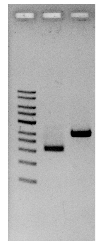
Plasmid DNA were loaded on 1% of agarose gel after extraction.
Comparison of workflows of Magnetic Beads based kits
Features
100% centrifuge-free
Culture medium can be used directly without centrifuge to pellet the bacteria
Simple
No centrifuge
No column
No vacuum
Flexible applications with simplified miniprep protocol
High throughput: 96-well plates with 0.2 ml of each culture (Cat.# 50011)
Low throughput: 1.5 ml tubes with 0.2 ml of each culture (Cat.# 50011)
Low throughput: 1.5 ml tubes with 2 ml of each culture (Cat.# 50012)
One cDNA Synthesis, Multiple microRNAs and microRNA-targets analyzed
Time Savings
Cost Efficient
High Sensitivity and Yield
Robust Enzyme
Available in 12 or 50 reaction size
Norgen’s microScript microRNA cDNA Synthesis Kit is an all-in-one, ready-to-use product for the reverse transcription of microRNA from either Total RNA preparations or enriched microRNA preparations. The kit contains the 2x Reaction Mix and the microScript microRNA Enzyme Mix. The kit utilizes Norgen’s microScript Reverse Transcriptase, a mutant version of Moloney Murine Leukemia Virus (M-MuLV) Reverse Transcriptase. It has reduced RNase H activity and increased thermal stability.
The workflow of Norgen’s microScript microRNA cDNA Synthesis Kit involves a simple, single-tube set-up by the mixing of 2x Reaction Mix, Enzyme Mix and the RNA template. The reaction can then be carried out in a thermocycler. A poly (A) tail is first added to the RNA template, followed by cDNA synthesis using an adapter primer. In addition to the ease-of-use, the single-tube set-up provides superb consistency and sensitivity. The cDNA could be used in a PCR or qPCR amplification using a Universal PCR Reverse Primer and the forward primer that contains the sequence of the microRNA of interest. A single cDNA preparation could be used for PCR amplification of a number of different microRNAs. In addition, the cDNA preparation could be used for PCR or qPCR detection (using gene-specific forward and reverse primers) of mRNA or large RNA if total RNA preparation was the starting template. This could allow for parallel evaluation of expression level of microRNAs and microRNA-targets.
t-Boc-N-amido-PEG4-Amide-Tri-(propargyl-PEG10-ethoxymethyl)-methane is a branched crosslinker molecule with three terminal propargyl groups and a t-Boc protected amide group. The propargyl groups can react with azide-bearing compounds or biomolecules via copper catalyzed azide-alkyne Click Chemistry to yield a stable triazole linkage. The protected amine can be deprotected under acidic conditions. Reagent grade, for research use only.
Document
t-Boc-N-amido-PEG4-Amide-Tri-(propargyl-PEG10-ethoxymethyl)-methane is a branched crosslinker molecule with three terminal propargyl groups and a t-Boc protected amide group. The propargyl groups can react with azide-bearing compounds or biomolecules via copper catalyzed azide-alkyne Click Chemistry to yield a stable triazole linkage. The protected amine can be deprotected under acidic conditions. Reagent grade, for research use only.