
Description
Specifications
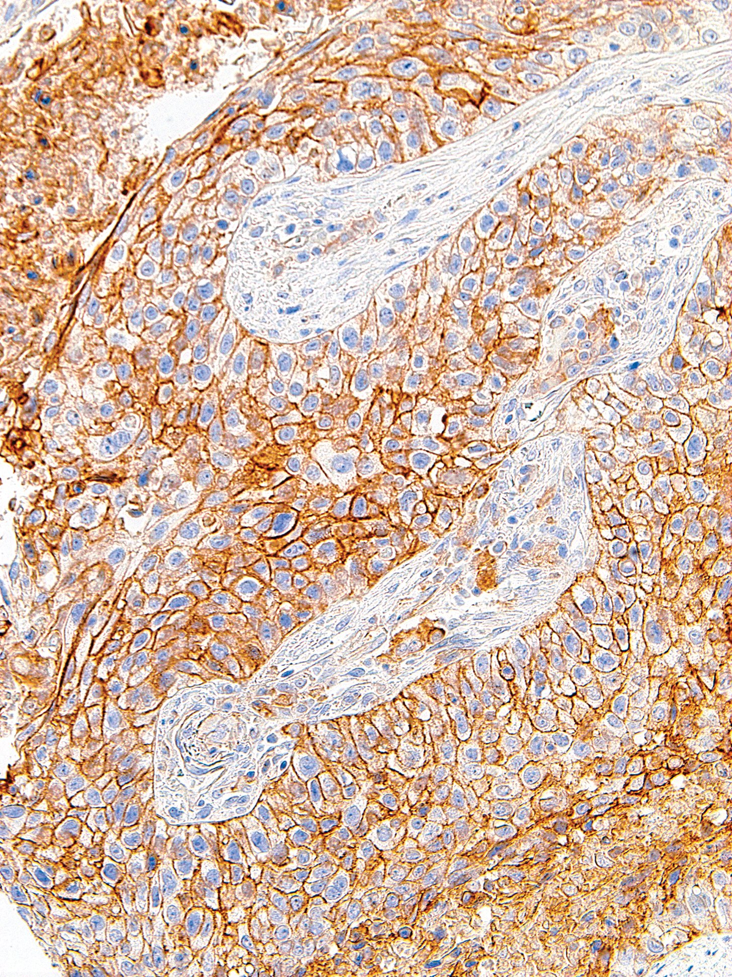
| Clone | IHC411 |
| Source | Rabbit Monoclonal |
| Positive Control | Tonsil, Lung Adenocarcinoma |
| Dilution Range | 1:200 |

| Clone | IHC411 |
| Source | Rabbit Monoclonal |
| Positive Control | Tonsil, Lung Adenocarcinoma |
| Dilution Range | 1:200 |

Lateral flow strips designed to detect a biotin and FITC/FAM labelled amplicon. Milenia Genline HybriDetect 1 (PDF 276 KB) lateral flow strips can be used to detect amplification products generated when using a TwistAmp® nfo kit in combination with a TwistAmp® nfo probe and labelled amplification primer.
Lateral flow strips designed to detect a biotin and FITC/FAM labelled amplicon. Milenia Genline HybriDetect 1 (PDF 276 KB) lateral flow strips can be used to detect amplification products generated when using a TwistAmp® nfo kit in combination with a TwistAmp® nfo probe and labelled amplification primer.

Product Details
Kit Size:
48 Reactions
Product Name:
DNA Isothermal Rapid Amplification Kit Fluorescent Type
Kit Type:
DNA
Reaction Volume:
50 μL
Storage Temperature:
-20℃
Application:
Nucleic Acid Amplification
Type:
Fluorescent Type
Reaction Time:
20mins
High Light:
,
,
Payment & Shipping Terms
Minimum Order Quantity
48T
Price
3.8$/T
Packaging Details
16T/bag,48T/box
Delivery Time
6days
Payment Terms
T/T,Paypal
Supply Ability
100000T/Month
Product Description
Product parameters:

| Reagent component ( WLE8202KIT ,16T/bags,48T/Box ) | |||
| Component | Specification | Quantity | Function |
| A buffer | 1.6ml | 1 Tube | Buffer system mainly for stabilizing protein/enzyme and performance |
| B buffer | 0.15ml | 1 Tube | Mainly activated systems such as magnesium ions |
| Positive control template | 0.1ml | 1 Tube | Mainly the positive plasmid template is used to test the effectiveness of the kit |
| Positive control primer mix | 0.06ml | 1 Tube | Mainly the primer combination of the positive control template |
| Reagent Guide Manua | 16T/bags,48T/Box | 3 bags | Reagent technology of protein/enzyme system: freeze-dried powder, freeze-dried microspheres |
Principle overview
This kit is based on a room temperature and constant temperature nucleic acid rapid amplification technology: at room temperature and constant temperature, the recombinase and primer form the protein/single-stranded nucleotide complex Rec/ssDNA, with the help of auxiliary proteins and single-stranded binding protein SSB , invade the double-stranded DNA template; form a D-loop region at the invasion site, and start scanning the DNA double-strands; after finding the target region complementary to the primer, the Rec/ssDNA complex disintegrates, and the polymerase also binds to The 3′ end of the primer initiates chain extension. This kit relies on the action of exonuclease at 39 ºC, adding specific molecular probes designed based on the template, and using fluorescence monitoring equipment to achieve real-time monitoring of the amplification process of the target fragment.
Primer design
It is recommended to use primers with a length of 30-35 bp. Primers that are too short will affect amplification speed and detection sensitivity; primers are designed to avoid the formation of secondary structures that affect amplification; the amplicon length is recommended to be 150-300 bp, usually no more than 500 bp.
Fluorescent probe design
The probe sequence does not overlap with the specific primer recognition site, is 46-52 nt in length, and the sequence avoids palindromic sequences, internal secondary structures, and continuous repeated bases. The probe has four modification sites: the middle position ≥ 35 nt from the 5′ end is labeled with a dSpacer (tetrahydrofuran, THF) as the recognition site for exonuclease; the upstream of the THF site is labeled with a fluorescent group, and the downstream Label a quenching group, the distance between the two groups is 2-4 nt; THF is ≥15 nt from the 3′ end, and the 3′ end is labeled with a modifying group, such as an amine group, a phosphate group or a C3-Spacer.
Product features and advantages:
This kit has the advantages of high sensitivity, strong specificity,and short reaction time (only 20 minutes), and the reaction groups are in dry powder state, which is easy to operate and easy to store.
It can be applied to various brands of fluorescence quantitative PCR instruments, constant temperature fluorescence amplification instruments and other fluorescence detection equipment.
This kit is based on a room temperature and constant temperature nucleic acid rapid amplification technology: at room temperature and constant temperature, the recombinase and primer form the protein/single-stranded nucleotide complex Rec/ssDNA, with the help of auxiliary proteins and single-stranded binding protein SSB , invade the double-stranded DNA template; form a D-loop region at the invasion site, and start scanning the DNA double-strands; after finding the target region complementary to the primer, the Rec/ssDNA complex disintegrates, and the polymerase also binds to The 3′ end of the primer initiates chain extension. This kit relies on the action of exonuclease at 39 ºC, adding specific molecular probes designed based on the template, and using fluorescence monitoring equipment to achieve real-time monitoring of the amplification process of the target fragment.
