5 tests Lateral Flow Assay designed for the detection of Human Anti Mouse Antibodies (HAMA) in human serum.
Detail
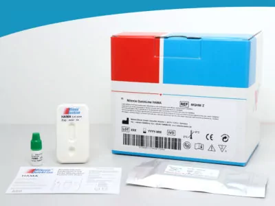
Name of Product
HAMA – Human Anti Mouse Antibodies Detection Kit
Catalog Number
MQHM Z
Short Info
Lateral Flow Assay designed for the detection of Human Anti Mouse Antibodies (HAMA) in human serum.
This product is only available in the EU!
Method/Platform
Lateral Flow
Range/Assay Sensivity
97%
Test Principle
The HAMA Detection Kit is an immunoassay designed for qualitative determination of Human Anti Mouse Antibodies in human serum.
HAMA from patient’s sample bind first to mouse antibodies which are conjugated to gold nanoparticles.
In a second step the HAMA will be caught by mouse IgG antibodies, coated on the Test Line (T) of the nitrocellulose membrane.
In the presence of the HAMA-conjugat-complex a band becomes visible at the location of the Test Line (T).
The surplus of gold particles continues to migrate through the membrane and is captured at the Control Line (C) by specific antibodies.
Color’s intensitiy of the test line is directly proportional to the HAMA concentration in the sample.
Brief Instructions
Storage
2-8°C
Components
HAMA Test Unit, Chase Buffer, Evaluation Cards
Other Products
IVD5116 HiPure FFPE DNA/RNA Kit
Product Info
Document
Product Info
Introduction
The Kit is specially designed for simultaneous purification of genomic DNA and total RNA from formalin-fixed, paraffin-embedded (FFPE) tissue sections. The Purified DNA/RNA is used for RT-PCR and PCR detection.
Details
Specifications
Features
Specifications
Main FunctionsC
Co-isolation DNA and RNA from a single FFPE tissue sample
Applications
RT-PCR, cDNA synthesis, PCR and second-generation sequencing, etc.
Purification method
Mini spin column
Purification technology
Silica technology
Process method
Manual (centrifugation or vacuum)
Sample type
FFPE slice, FFPE embedded tissue
Sample amount
No more than six 10µm sections of 150mm2 surface area or three 20µm sections of 150mm2 surface area.
Principle
FFPE samples are incubated in an optimized lysis buffer, which results in the release of RNA and precipitation of DNA. After centrifugation, the RNA-containing supernatant and DNA-containing pellet are then processed separately to purify RNA and DNA. For RNA purification, transfer RNA Lysate to an adsorption column and RNA is adsorbed on the membrane, while protein is not adsorbed and is removed with filtration. After washing proteins and other impurities, RNA was finally eluted with low-salt buffer. For DNA purification, transfer DNA Lysate to an adsorption column and DNA is adsorbed on the membrane, while protein is not adsorbed and is removed with filtration. After washing proteins and other impurities, DNA was finally eluted with low-salt buffer.
Advantages
1.Use non-toxic dewaxing solution without contact with xylene 2.Obtain both DNA and RNA simultaneously from the same sample. Elute separately without affecting each other (Have the same steps and effects as top brand 80234, perfect substitute.)
Kit Contents
Contents
IVD5116
Purification Times
50 Preps
HiPure DNA Micro Column
50
HiPure RNA Mini Column I
50
2ml Collection Tubes
150
Proteinase K
50 mg
Protease Dissolve Buffer
5 ml
Buffer DPS
60 ml
Buffer FRL
15 ml
Buffer ATL
15 ml
Buffer RLC
15 ml
Buffer AL
15 ml
Buffer VHB
44 ml
Buffer RW2
25 ml
RNase Free Water
10 ml
Buffer AE
10 ml
Storage and Stability
Proteinase K should be stored at 2-8°C upon arrival. However, short-term storage (up to 12 weeks) at room temperature (15-25°C) does not affect their performance. The remaining kit components can be stored at room temperature (15-25°C) and are stable for at least 18 months under these conditions.
Experiment Data
Document
The Kit is specially designed for simultaneous purification of genomic DNA and total RNA from formalin-fixed, paraffin-embedded (FFPE) tissue sections. The Purified DNA/RNA is used for RT-PCR and PCR detection.
Propargyl-PEG4-sulfonic acid is heterobifunction reagent that can reacts with azide compounds or biomolecules via copper catalyzed Click Chemistry reactions. Reagent grade, for research purpose. Please contact us for GMP-grade inquiries.
Document
Propargyl-PEG4-sulfonic acid is heterobifunction reagent that can reacts with azide compounds or biomolecules via copper catalyzed Click Chemistry reactions. Reagent grade, for research purpose. Please contact us for GMP-grade inquiries.