
Introduction
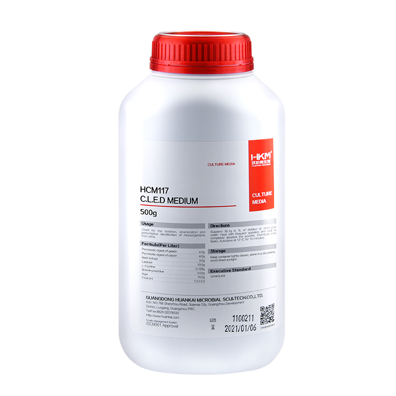
Usages: For the total microbial count in urine.
Storage: Keep container tightly closed, store in a cool, dry place, away from bright light. Storage period of 3 years.

Usages: For the total microbial count in urine.
Storage: Keep container tightly closed, store in a cool, dry place, away from bright light. Storage period of 3 years.


General information
Champion™ Competent Cells are chemically competent cells, which were prepared by SMOBIO to make E. coli perform excellent transformation efficiency. Standard transformation protocol is recommended for large plasmids or non-ampicillin selection. Time-saving transformation protocol is recommended for simple and rapid transformation. Champion™ Competent Cells are one of the fastest and simplest ready-to-use competent cell products in the world.
Kit contents
Shipping condition
Throughout the shipping process, the temperature is maintained under -70°C.
Storage and expiration
Champion™ Competent Cells must be stored between -70°C to -80°C. Subsequent freeze-thaw cycles will reduce transformation efficiency. If high efficiency is required for the experiment, do not use aliquots that have gone through several freeze-thaw cycles. The efficiency of Champion™ Competent Cells lasts for 1 year with proper storage.
Champion™ Competent Cells are chemically competent cells, which were prepared by SMOBIO to make E. coli perform excellent transformation efficiency. Standard transformation protocol is recommended for large plasmids or non-ampicillin selection. Time-saving transformation protocol is recommended for simple and rapid transformation. Champion™ Competent Cells are one of the fastest and simplest ready-to-use competent cell products in the world.

Permagen’s most universal ring magnet plate. From Low elution PCR applications up to 2 mL deep well, the X96 has you covered. No need to purchase two separate plates anymore. Smaller inner ring magnet allows for volumes as low as 5 µL (from PCR plates), larger outer magnet handles up to 2 mL deep well assays
ANSI/ SBS Footprint (127.75mm x 85.50mm) to fit into any automated liquid handling robot on bottom, SBS/ SLAS fit on top to accept any microplate
Integrated Cushion base for maximum recovery. Helps aid in set-up, robot positioning inconsistencies, and labware consumable differences
Features include solid aluminum alloy construction and hard coat anodized finish for years of trouble-free use, and compatible with any magnetic beads
A011623
A011623
Maximum – 2 mL (deep well plate
Minimum – 5 µL (pcr plate, or tubes with kit)
Permagen’s most universal ring magnet plate. From Low elution PCR applications up to 2 mL deep well, the X96 has you covered. No need to purchase two separate plates anymore. Smaller inner ring magnet allows for volumes as low as 5 µL (from PCR plates), larger outer magnet handles up to 2 mL deep well assays