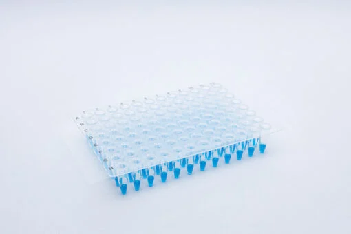
A transparent, optically clear, DMSO resistant, non-tacky film which adheres only when pressure is applied. It is non-pierceable and peelable. Clear plastic, reflective, glossy on the top. Very thin and light and doesn’t crease easily.
Detail
Overview
A transparent, optically clear, DMSO resistant, non-tacky film which adheres only when pressure is applied. It is non-pierceable and peelable. Clear plastic, reflective, glossy on the top. Very thin and light and doesn’t crease easily.
Crystal clear seal specifically developed for optical applications, particularly qPCR
Non-sticky when removed from the packaging aiding with handling when using gloves
When the seal is in position, pressure can be applied to burst the capsules, releasing a strong adhesive only where the seal touches the raised well rims of the plate – the rest of the seal area above the wells remains optically clear
Other Products
[PM2700] ExcelBand™ 3-color Broad Range Protein Marker (3.5-245 kDa), 250 μl x 2
Product Info
Document
Product Info
Description
The PM2700 ExcelBand™ 3-color Broad Range Protein Marker is a ready-to-use three-color protein standard with 13 pre-stained proteins covering a wide range of molecular weights from 5 to 245 kDa in Tris-Glycine buffer (3.5 kDa to 235 kDa in Bis-Tris (MOPS) buffer and Bis-Tris (MES) buffer). Proteins are covalently coupled with a blue chromophore except for two reference bands (one green and one red band at 25 kDa and 75 kDa, respectively) when separated on SDS-PAGE (Tris-Glycine buffer). The PM2700 ExcelBand™ 3-color Broad Range Protein Marker is designed for monitoring protein separation during SDS-polyacrylamide gel electrophoresis, verification of Western transfer efficiency on membranes (PVDF, nylon, or nitrocellulose) and for approximating the size of proteins.
Features
Ready-to-use — Premixed with a loading buffer for direct loading, no need to boil.
Two reference bands — 75 kDa (red) and 25 kDa (green)
Contents
Approximately 0.1~0.4 mg/ml of each protein in the buffer (20 mM Tris-phosphate (pH 7.5 at 25°C), 2% SDS, 0.2 mM DTT, 3.6 M urea, and 15% (v/v) glycerol).
Quality Control
Under suggested conditions, PM2700 ExcelBand™ 3-color Broad Range Protein Marker resolves 13 major bands in SDS-PAGE (Tris-Glycine buffer, MOPS, and MES buffer) and after Western blotting to nitrocellulose membrane.
Storage
4°C for 3 months -20°C for long term storage
Document
The PM2700 ExcelBand™ 3-color Broad Range Protein Marker is a ready-to-use three-color protein standard with 13 pre-stained proteins covering a wide range of molecular weights from 5 to 245 kDa in Tris-Glycine buffer (3.5 kDa to 235 kDa in Bis-Tris (MOPS) buffer and Bis-Tris (MES) buffer). Proteins are covalently coupled with a blue chromophore except for two reference bands (one green and one red band at 25 kDa and 75 kDa, respectively) when separated on SDS-PAGE (Tris-Glycine buffer). The PM2700 ExcelBand™ 3-color Broad Range Protein Marker is designed for monitoring protein separation during SDS-polyacrylamide gel electrophoresis, verification of Western transfer efficiency on membranes (PVDF, nylon, or nitrocellulose) and for approximating the size of proteins.
Short term stability: 2-8oC, Long term stability: See individual component labels
Stability:
> 2 years under recommended storage conditions
Analyte:
D-Glucose, Raffinose, Sucrose
Assay Format:
Spectrophotometer
Detection Method:
Absorbance
Wavelength (nm):
510
Signal Response:
Increase
Limit of Detection:
100 mg/L
Reaction Time (min):
~ 20 min
Application examples:
Analysis of grain legumes and other materials containing raffinose, stachyose and verbascose.
Method recognition:
Used and accepted in food analysis
The Raffinose/Sucrose/D-Glucose test kit is for the measurement and analysis of D-glucose, sucrose and raffinose, stachyose and verbascose in seeds and seed meals. Based on the measurement of D-glucose on enzymic hydrolysis of raffinose, stachyose and verbascose to D-glucose, D-fructose and D-galactose.
All reagents stable for > 2 years after preparation
Simple format
Rapid reaction
Mega-Calc™ software tool is available from our website for hassle-free raw data processing
Standard included
Document
The Raffinose/Sucrose/D-Glucose test kit is for the measurement and analysis of D-glucose, sucrose and raffinose, stachyose and verbascose in seeds and seed meals. Based on the measurement of D-glucose on enzymic hydrolysis of raffinose, stachyose and verbascose to D-glucose, D-fructose and D-galactose.
KBA.62, also known as Melanoma Associated Antigen, is used to detect an antigen present in melanocytic tumors, such as melanomas, due to its proven sensitivity and specificity. The antibody can also be used to distinguish between junctional nevus and intradermal nevus cells, and fetal melanocytes versus normal adult melanocytes. Studies have shown KBA.62 to be highly useful in differentiating between metastatic amelanotic melanoma and a number of poorly differentiated carcinomas, large cell lymphomas, sarcomas, and spindle cell carcinomas.